|
#1
●
05-02-2024, 04:57 PM
| ||||||||
 These are the rooms Poster Rank:24 of ruin. Join Date: Sep 2014 Posts: 54,011 Mentioned: 145 Post(s) Quoted: 30403 Post(s)
| ||||||||
|
Avulsion Of Facial Skin
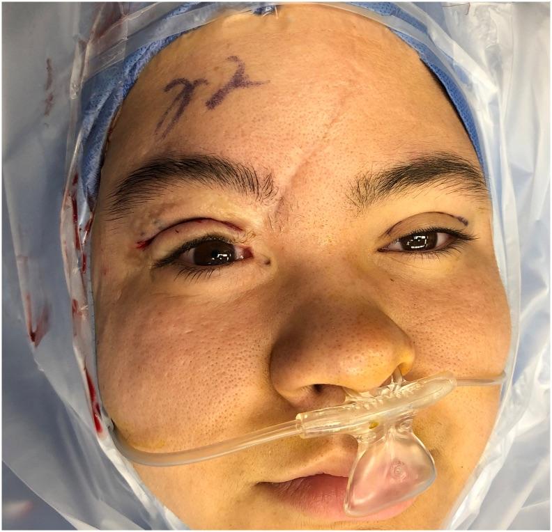
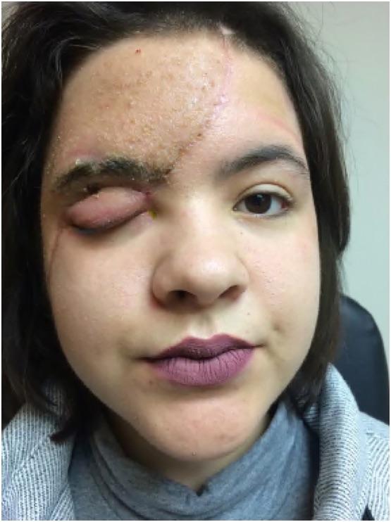
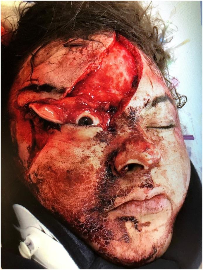
A 19-year-old female was the restrained passenger in a motor vehicle accident brought to a Level 1 Trauma Center for evaluation and treatment. Initial evaluation of the patient revealed complete avulsion of the tissues of the right cheek, upper eyelid, and forehead (Fig. 1). Primary repair of the cheek, eyelid, and forehead wounds was performed. The repair of the eyelid wound was done without exploration or reinsertion of the LPSA. After the repair, oculoplastics was consulted for evaluation of complete right upper eyelid ptosis. >>External exam revealed significant edema of the right upper eyelid, with no eyelid crease present. There was right brow ptosis, with the patient unable to activate the frontalis on the right with normal contraction on the left. There was complete ptosis of the right upper eyelid (margin to reflex distance (MRD1)) −3.0 mm, vertical fissure (VF) 0 mm, with a levator function of 0 mm. The left upper eyelid had a MRD1 of 3.0 mm, VF of 8.0 mm, and levator function of 13 mm. >>At the one-month follow up, the patient continued to have complete ptosis with no levator function (Fig. 2). Right brow ptosis and frontalis paresis was still present. The patient returned for a six-month follow up and continued to have complete right upper eyelid ptosis with no LF, brow ptosis, and frontalis paresis. Exploratory surgery was planned for the following week, but did not occur due to difficulty with patient scheduling. The patient returned three months later with improvement of the ptosis and levator function, but no improvement of brow ptosis or frontalis function. In the right eye, MRD1 was 2 mm with a vertical fissure of 7 mm. The left eye MRD1 was 4 mm and vertical fissure 9 mm. The levator function was 8 mm on the right and 13–14 mm on the left. The patient was concerned about the contour and position of the right upper eyelid compared to the left. External levator advancement and exploration was performed the following week with good intraoperative results (Fig. 3, Video 1). The patient gave informed consent for images and video of relating to her case to be used for lectures and/or publication. |
|
#3
●
05-02-2024, 09:38 PM
| ||||||||
 ★ Legacy Member ★ Poster Rank:111 Secret Agent Join Date: Dec 2009 Posts: 14,328 Mentioned: 6 Post(s) Quoted: 2980 Post(s)
| ||||||||
|
Re: Avulsion Of Facial Skin
They did pretty good considering that bloody photo.  She was pretty wrecked. Now she’s pretty cute. She needs a pirate patch over the right eye though and maybe some swashbuckling boots.  |
|
#4
●
05-02-2024, 10:45 PM
| ||||||||
 ♚ Legacy Gold Member ♚ Poster Rank:265 ꓕSIꓤIꓕ∀S ϽINOᗡꓤ∀S Join Date: Feb 2010 Posts: 5,156
Contributions: 6
Mentioned: 42 Post(s) Quoted: 1435 Post(s)
| ||||||||
|
Re: Avulsion Of Facial Skin
She can thank the miracle of super glue... and long bangs.
|
|
#5
●
05-03-2024, 10:00 PM
| ||||||||
 My Rank: LANCE CORPORAL Poster Rank:1848 Male but Gay and if someone Hate´s me: Excuse me ich havent Choose it freely. Join Date: May 2017 Posts: 278 Mentioned: 1 Post(s) Quoted: 79 Post(s)
| ||||||||
|
Re: Avulsion Of Facial Skin
Nobody knows that in the Internal Medicine Cyanacrylat (the main Substance from Superglue) is used to glue Holes in the Heart or other Internal Organs. Many People where not alive if the don had the Superglue.  |