|
#1
●
05-29-2024, 10:44 PM
| ||||||||
 These are the rooms Poster Rank:26 of ruin. Join Date: Sep 2014 Posts: 54,011 Mentioned: 145 Post(s) Quoted: 30403 Post(s)
| ||||||||
|
Attacked With A Plank
This was a 62-year-old man with type 2 diabetes who was assaulted with a wooden plank and rusty nail on his frontal scalp area sustaining a laceration 3 days prior. The laceration was sutured and a head injury was ruled out with a CT scan. The next day, he developed a rapid progressive bilateral periorbital cellulitis and proptosis followed by transient loss of vision of his right eye (Fig. 10). He was pyrexial at 38.7 °C and confused. WCC was 24.5 × 10⁹/L and CRP was 330 mg/L. He was immediately started on meropenem and clindamycin. Repeat CT orbit showed extensive bilateral periorbital swelling with stranding of the subcutaneous fat with fluid collections within the pretarsal regions and lateral to the globes. There was extensive soft tissue swelling over the nasal bridge (Fig. 11). He underwent immediate debridement of the necrotic tissues. This included the underlying fascia, frontalis, and nasal bridge skin to the lid margin, including the orbicularis oculi which was resected. Orbital septum and levators were left intact (Fig. 12). The extent of the defects was similar bilaterally and extended to the vertex with bare periosteum in the midline (Fig. 13). A week later, reconstruction was performed. Scalp flaps were raised bilaterally (Fig. 14). Both cheeks were advanced superiorly for lower eyelid reconstruction using nasolabial incisions (Fig. 15). A lateral canthopexy was performed bilaterally. Palmaris tendon was used as slings for lower eyelid support bilaterally (Fig. 16). The upper eyelids were reconstructed using compound grafts from the scrotum incorporating skin and cremasteric muscle. Sural nerve grafts were coapted to the frontal nerve bilaterally (Fig. 17) and directly onto the cremasteric muscular aspect of the compound graft to neuratize and reinnervate this muscle for upper eyelid dynamic reconstruction in both upper eyelids (Fig. 18). The cremasteric muscle is a thin layer of striated muscle found in the scrotum between the external and internal layers of spermatic fascia that functions to raise and lower the testes for temperature regulation. It demonstrates a cremasteric reflex as well as contracts voluntarily. It is supplied by the cremasteric artery, a branch of the inferior epigastric artery and the genitofemoral nerve, both of which were not dissected out. Instead, this muscle was neuratized by the frontal nerve and sural nerve graft. This was based on the concept of muscle graft for dynamic facial reanimation first described by Noel Thompson in 1971 [4] and also Nassif et al. [5] The reason for using the scrotum as a donor site for the skin graft, along with the cremasteric muscle, was twofold. Firstly, as skin grafts were required for the upper eyelid reconstruction, a compound graft with the cremasteric muscle component would serve the secondary purpose of dynamic reconstruction for spontaneous eye closure. Secondly, with the frontal nerve identified and readily available post-debridement, coapting a nerve graft for the short distance to the muscle graft on each side seems an obvious reconstructive step. Further conventional technique, albeit more complex and time-consuming surgery using the platysma muscle to restore spontaneous blink, would still be possible by re-coaptation to the same nerve graft in the future and tunneling the muscle under the previous graft, so no bridges were burnt. With further lateral canthopexy procedures 6 months down the line, the patient could achieve closure of his eyelids even though spontaneous blink was not present. However, the patient declined further surgery and was satisfied with the result having had no episodes of eye infections. 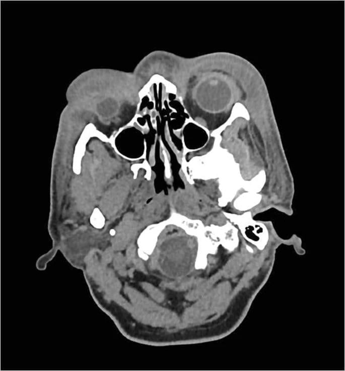 ·  · 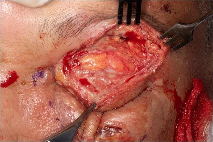 · 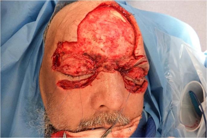 · 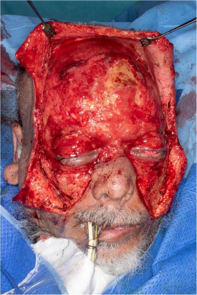 · 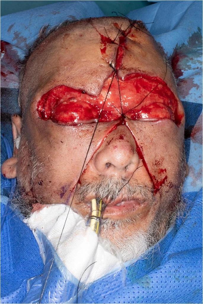 ·  · 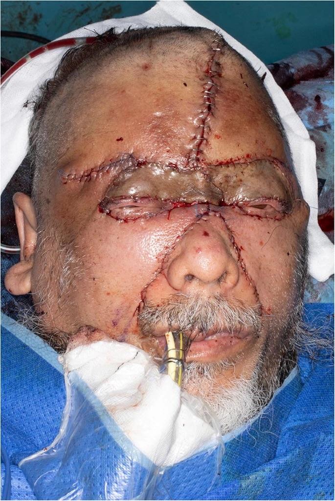 ·  · |
|
#3
●
05-30-2024, 01:00 AM
| ||||||||
 My Rank: FIRST SERGEANT Poster Rank:410 Male Join Date: Nov 2009 Posts: 2,968 Mentioned: 18 Post(s) Quoted: 448 Post(s)
| ||||||||
|
Re: Attacked With A Plank
Flaps of scrotum for eyelids... Nifty trick. |