|
#1
●
08-15-2024, 10:42 PM
|
 These are the rooms Poster Rank:26 of ruin. Join Date: Sep 2014 Posts: 54,008 54,008Posts 87,658Thanked 30,403Quoted 145Mentioned 0Contrib. |
|
Ankylosing Spondylitis and Kyphosis Deformity
This study reported a patient, male, 33 years old, mongoloid, working as an administration employee, without any history of smoking or alcohol consumption. Who was diagnosed with Ankylosing Spondylitis and Kyphosis deformity. Patient had history of slouching back from age of 18 years old with no history of trauma nor family history. The slouching back got worse as the patient got older, with new onset of back pain in the age of 33 years old. Patient got examined by a Spine Orthopaedic surgeon, from the lab workup there was no abnormality, and the x-ray examination revealed a severe kyphotic deformity with ankylosing of the spine. Patient was diagnosed as Ankylosing Spondylitis and Kyphosis and underwent deformity correction surgery (Fig. 1, Fig. 2). The post-operative outcome was excellent with no wound dehiscence for 5 weeks follow up in ward. Patient was discharged from the hospital and took oral antibiotic for 1 week; patient had no history of allergy for the medication. Patient should had come every week after the discharge, instead he came 1 month later complained about his open surgical wound with the size of 30 × 15 × 3 cm3 surrounded with necrotic tissue with bone as the base. In our institution, this patient underwent second debridement surgery and unilateral reverse latissimus dorsi flap for thoracal soft tissue defect coverage to obtain well-vascularized tissue, with good resistance to bacterial contamination and easy to shape into such defect. Before surgery, the patient should achieve the Hb level of minimal 10 g/dl, Albumin level of minimal 3.5 g/dl. The preoperative antibiotic used was Intravenous Cefazoline 2 g. For the surgical procedure. The surgery was conducted by a Hand and Microsurgery Orthopaedic surgeon in a Siloam Hospital at Yogyakarta. First, we drew template on the patient. We drew the anatomical landmark such as contralateral scapula, midline, level of spine, and we also measured the size of the defect and made flap template in the contralateral axilla in oblique manner. We incised the flap template to expose the latissimus dorsi muscle. After that we raised the skin flaps and cut the latissimus dorsi muscle 10 cm from the insertion. We identified the thoracodorsal vessels and nerve, and we raised the muscle until the perforator from lumbar and posterior intercostal vessels. Then the flap was turned over under the subcutaneous tissue over the defect. After that the flap was attached with absorbable sutures. The donor site was sutured in layers and a drain was placed. After surgery patients was positioned in prone or lateral position, the drain was removed by 5 days and sutures were removed by 15 days. There were no complications during the surgery. The patient was given oral antibiotic for 1 week after discharged form hospital, the patient came to the outpatient clinic twice a week to evaluate the flap and the donor site, and the patient were asked to sleep in the prone or lateral position until the sutures were removed. Patient followed all the postoperative instruction. The outcome was evaluated 2 weeks after surgery. After follow-up for 6 months, the patient showed good outcome and postoperative wound healing was favorable. The flap was healed well. There was no any complication of the flap. 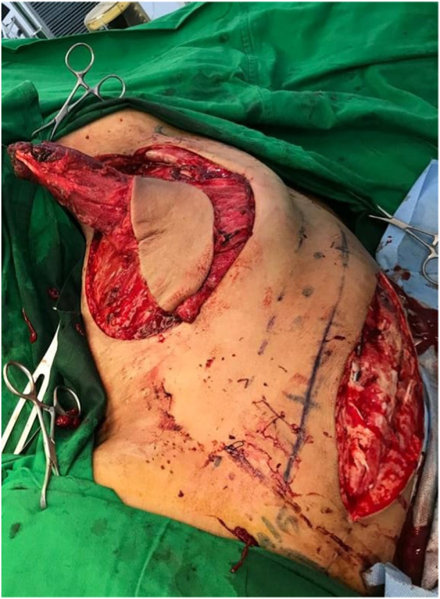 · 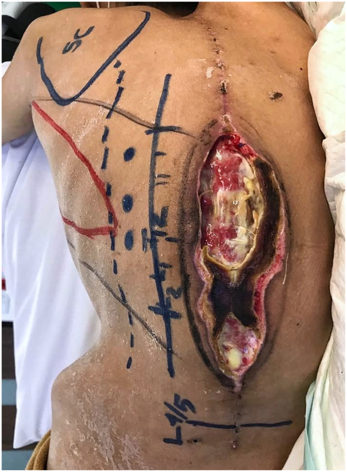 · 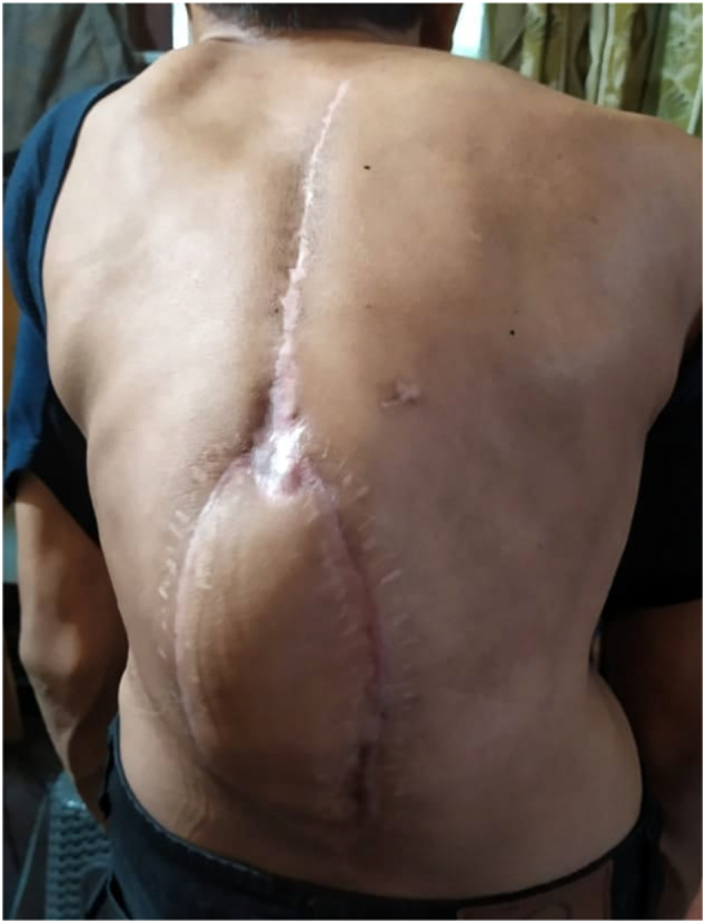 · |
|
#3
●
08-16-2024, 10:28 AM
|
 ✝Mudderator from Hell✝ Poster Rank:10 e-mail Join Date: Oct 2006 Posts: 96,690
Contributions: 901
96,690Posts 543,635Thanked 10,371Quoted 479Mentioned 901Contrib. |
|
Re: Ankylosing Spondylitis and Kyphosis Deformity
The "Rugby ball".  |