|
#1
●
07-01-2024, 09:23 PM
|
 These are the rooms Poster Rank:26 of ruin. Join Date: Sep 2014 Posts: 54,008 54,008Posts 87,658Thanked 30,403Quoted 145Mentioned 0Contrib. |
|
Ameloblastoma of the Mandible
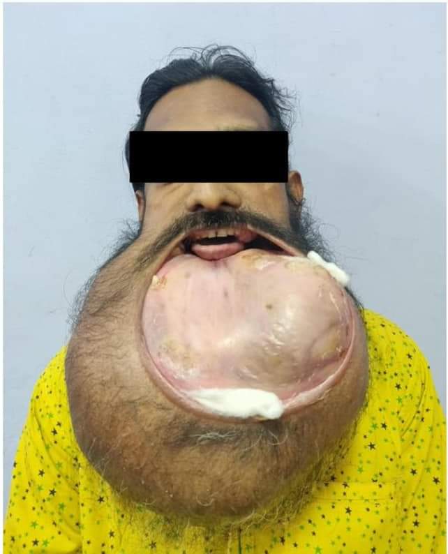
A 38-year-old, thin-built gentleman was brought by our hospital’s community outreach unit with a gradually progressive, painless swelling of the lower jaw for ten years. As it increased to the size of a football, he sought medical help, however, in view of the enormous size and other medical issues, surgery was deferred at various other centers. Social stigma and financial constraints had gradually pushed him into clinical depression. Before coming to us, he had also taken native medicinal treatment for a short duration. Clinical examination revealed a large, non-tender swelling with variable consistency measuring 25×22 cm involving the entire body of the mandible, up to the angles bilaterally, with overtly stretched lower lip and overlying skin. Intra-orally, floor of the mouth and lower gingivobuccal sulcus too were significantly stretched with loss of multiple teeth from the lower jaw, with few displaced residual teeth still attached to the distorted mandible with no obvious ulcer/lesions in the oral cavity. He had a fair mouth opening, with no restriction of tongue movements and was able to consume a soft diet (Figure 1). Evaluation with a three-dimensional CT of head and neck (Figure 2), revealed a huge expansile lytic lesion of size 25×22×15 cm involving the entire body of the mandible, with overtly stretched floor of mouth muscles, with loss of fat planes. The expanded mandibular cortex was partly thickened and partly invisible and had internal multilocular cystic and few solid enhancing components; features suggestive of a giant Ameloblastoma of the mandible. Work-up revealed low hemoglobin, thrombocytosis, hypertension, and a raised creatinine level. On further evaluation, in consultation with a hematologist, the anemia was found to be nutritional, for which he received blood transfusion prior to the surgery. No obvious cause for the thrombocytosis was identified. A physician's consult was sought, and he was started on an antihypertensive. The raised serum creatinine was thought to be pre-renal or secondary to the native medicines taken by the patient, however, it was corrected with adequate fluid resuscitation. A multi-disciplinary team meeting, involving the surgical oncologist, plastic and reconstructive surgeons, radiation oncologist and radiologist was held. A preoperative biopsy was deferred in view of the extensive lytic nature of the tumor and very few significant solid areas that could be targeted for the biopsy. As the entire body of mandible was involved and the CT imagining could not delineate the floor of mouth muscles clearly, prophylactic tracheostomy for adequate airway protection was planned. After ensuring adequate preoperative optimization, he was taken up for surgery i.e., wide local excision of the mandibular tumor and, single stage reconstruction with free fibula osteocutaneous flap. Due to the enormous size of the tumor, mask ventilation and laryngoscopy was difficult, hence an awake fiber optic nasal intubation was done under an airway block. Simultaneous mandibular resection and fibula free flap harvest were done by two teams. The gigantic size of the tumor had led to an altered local anatomy along with large, dilated vessels, which made resection of the tumor a challenging task. The entire body of the mandible involved by the tumor was resected, ensuring an adequate margin on either side. The harvest of fibula osteocutaneous flap was performed in the standard manner based on peroneal vessels from the left lower limb, i.e, 6 cm of fibula bone was preserved proximally to safeguard the Common peroneal nerve and 6 cm of bone was preserved distally to maintain ankle stability. Around 24 cm of segment of bone was safely harvested to obtain adequate pedicle length. Once harvested, multiple osteotomies were performed as per requirement i.e., 2 cm of bone for reconstruction of each ramus and 7 cm for the central body of mandible and plating was done (Figure 3). Refashioned fibula was then anchored to bilateral rami of the mandible using titanium plates and screws while maintaining an adequate maxillo-mandibular relationship and the skin paddle was used to drape the lingual surface of the bone (Figure 4). Microvascular anastomosis was performed between the peroneal artery and the facial artery, and the two venae comitantes were anastamosed with the Internal Jugular vein. Excess local soft tissue was excised and plantaris tendon was used as a static sling to maintain oral competence. Covering tracheostomy was done for airway protection, in view of extensive mandibular and floor of mouth resection, at the end of the surgery. The patient was monitored in the intensive care unit in the immediate postoperative period which was uneventful. He received nasogastric tube feeds in the immediate postoperative period and was then started on oral liquid feeds on postoperative day 10 which he tolerated well. Tracheostomy was decannulated once the surgical site edema resolved and after ensuring adequate tongue movements and swallowing reflexes. He was discharged from the hospital on postoperative day 13. Final histopathology reported a gross tumor weighing 5.4 kg, (Figure 5) measuring 25×20×16 cm, cut section showing a multicystic lesion with expanded mandibular bone with focal solid areas (Figure 6) and histology confirmed a conventional multicystic ameloblastoma (Figure 7) with reactive regional nodes with 2.5 cm margin. At four months follow-up, good oral competence and restored mandibular shape were observed. There is no sign of recurrence. As a tertiary care, not-for-profit centre, we were able to help the patient in a holistic manner, both medically and financially. The whole cost of hospitalization including the cost of implants was borne by the institute with the help of donors. With additional financial aid, we hope to aid in dental rehabilitation with osseo-integrated implants or dental prostheses in the future. 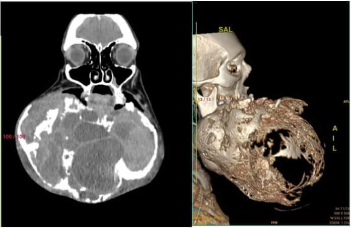 · 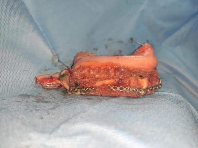 · 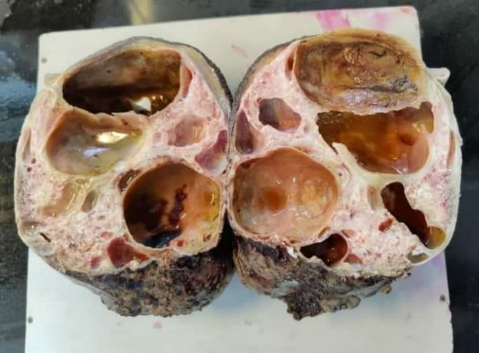 · 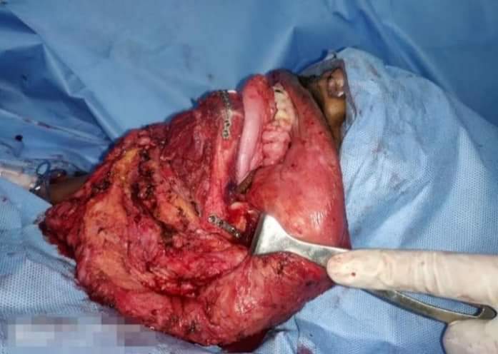 · 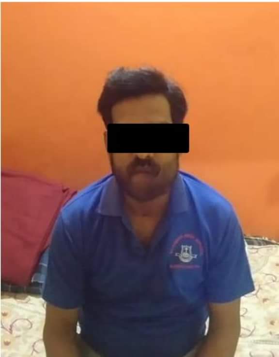 · |
|
#2
●
07-01-2024, 09:23 PM
|
| These are the rooms Poster Rank:26 of ruin. Join Date: Sep 2014 Posts: 54,008 54,008Posts 87,658Thanked 30,403Quoted 145Mentioned 0Contrib. |
|
Re: Ameloblastoma of the Mandible
Oops, forgot to add this.
 · |
|
#3
●
07-01-2024, 11:44 PM
|
 ★ Legacy Member ★ Poster Rank:252 So many choices now Join Date: Jul 2015 Posts: 5,542 5,542Posts 19,785Thanked 2,120Quoted 15Mentioned 0Contrib. |
|
Re: Ameloblastoma of the Mandible
That makes things a bit clearer.
|
|
#4
●
07-02-2024, 12:44 AM
|
 My Rank: MASTER GUNNERY SERGEANT Poster Rank:352 Manly Man Join Date: Sep 2017 Posts: 3,601 3,601Posts 12,427Thanked 700Quoted 0Mentioned 0Contrib. |
|
Re: Ameloblastoma of the Mandible
Had it for 10 years before he got help?? Dumbass.
|
|
#6
●
07-02-2024, 09:53 AM
|
 ★ Legacy Member ★ Poster Rank:110 Secret Agent Join Date: Dec 2009 Posts: 14,783 14,783Posts 49,087Thanked 3,049Quoted 6Mentioned 0Contrib. |
|
Re: Ameloblastoma of the Mandible
That kinda reminds me of a kangaroo pouch with a baby roo inside.  |
|
#7
●
07-11-2024, 03:22 AM
|
 My Rank: STAFF SERGEANT Poster Rank:728 Male Join Date: Sep 2009 Posts: 1,134 1,134Posts 1,537Thanked 119Quoted 1Mentioned 0Contrib. |
|
Re: Ameloblastoma of the Mandible
That thing belongs in a medical museum. (The growth, not the man)
|