|
#1
●
05-15-2016, 08:02 AM
| ||||||||
| ✝Mudderator from Hell✝ Poster Rank:10 e-mail Join Date: Oct 2006 Posts: 95,044
Contributions: 817
Mentioned: 472 Post(s) Quoted: 10089 Post(s)
| ||||||||
|
Abdominal Aortic Aneurysms (AAA) Rupture
Abdominal aortic aneurysms (AAAs) are relatively common and are potentially life-threatening. Patients at greatest risk for AAA are men who are older than 65 years and have peripheral atherosclerotic vascular disease. AAAs are usually asymptomatic until they expand or rupture. An expanding AAA causes sudden, severe, and constant low back, flank, abdominal, or groin pain. Syncope may be the chief complaint, however, with pain less prominent. Most clinically significant AAAs are palpable upon routine physical examination. The presence of a pulsatile abdominal mass is virtually diagnostic but is found in fewer than half of all cases. 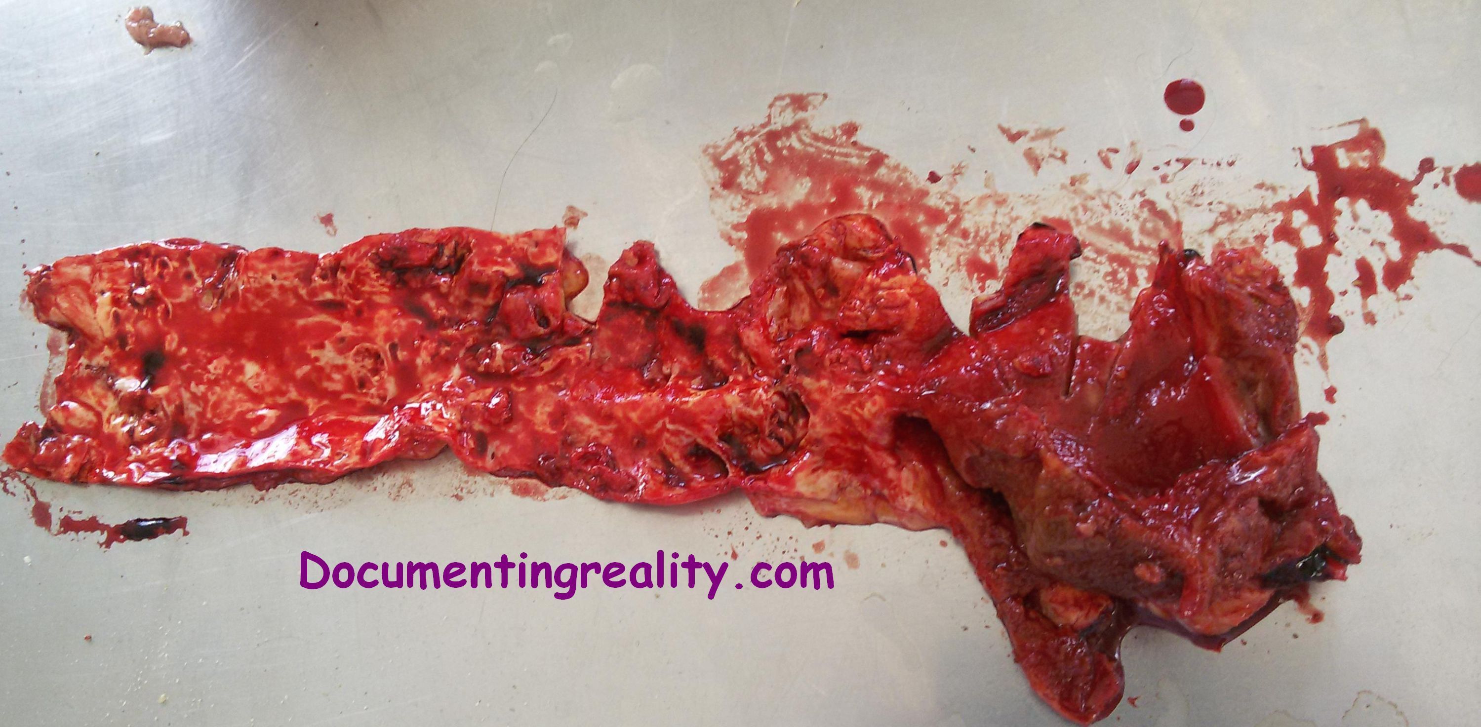 Patients with a ruptured AAA may present in frank shock, as evidenced by cyanosis, mottling, altered mental status, tachycardia, and hypotension. Whereas abrupt onset of pain due to rupture of an AAA may be quite dramatic, associated physical findings may be very subtle. Patients may have normal vital signs in the presence of a ruptured AAA as a consequence of retroperitoneal containment of hematoma. 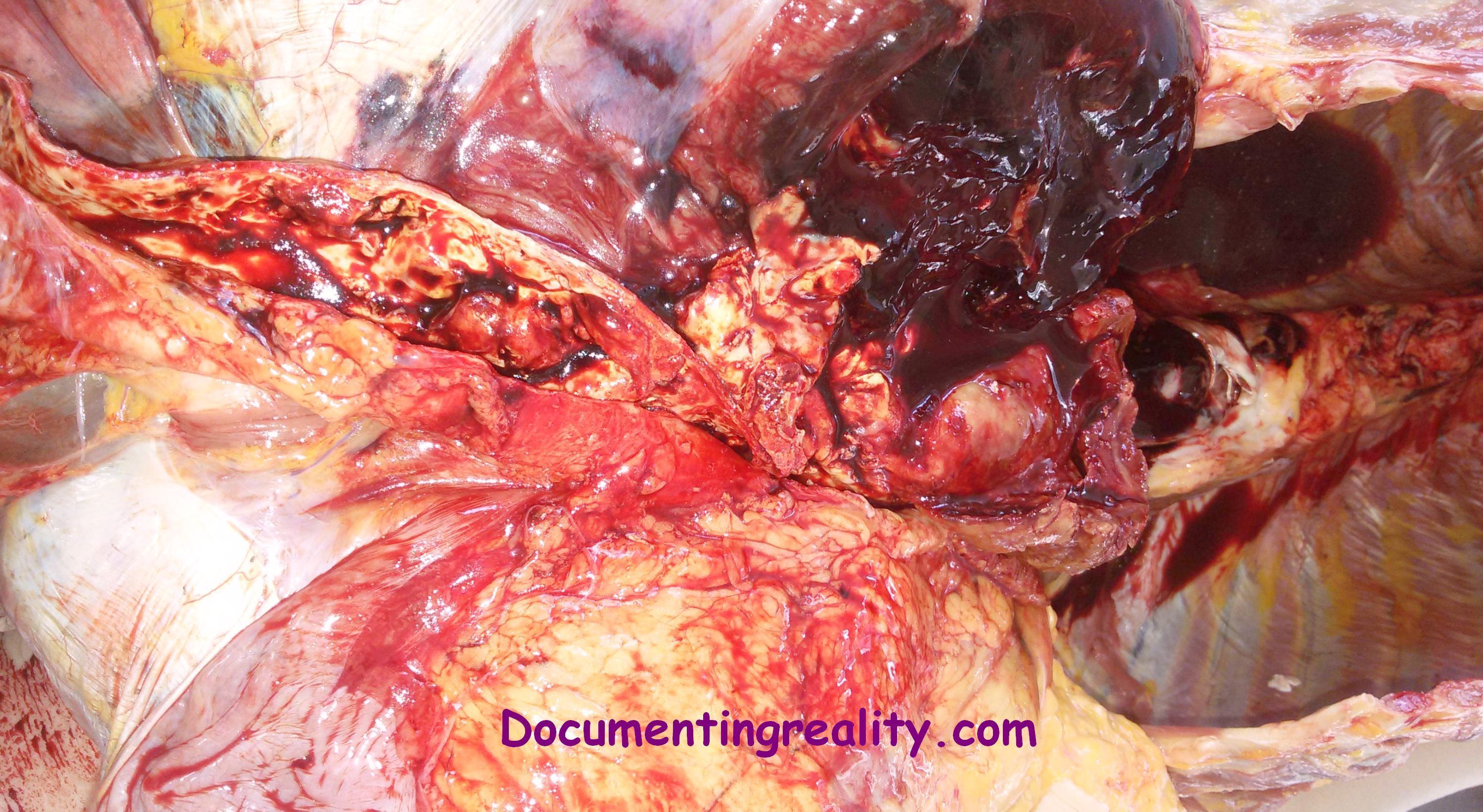 At least 65% of patients with a ruptured AAA die of sudden cardiovascular collapse before arriving at a hospital. Diagnosis : No specific laboratory studies can be used to diagnose AAA. The following imaging studies, however, can be employed diagnostically: Ultrasonography - Standard imaging technique for AAA Plain radiography - Using this method to evaluate patients with AAA is difficult because the only marginally specific finding, aortic wall calcification, is seen less than half of the time Computed tomography (CT) - This offers certain advantages over ultrasonography in defining aortic size, rostral-caudal extent, involvement of visceral arteries, and extension into the suprarenal aorta Magnetic resonance imaging - This permits imaging of the aorta comparable to that obtained with CT and ultrasonography, without subjecting the patient to dye load or ionizing radiation Angiography - This is helpful in determining aortic anatomy and has been advocated for preoperative use in certain cases Click pics for higher res. |
|
#3
●
05-16-2016, 04:09 AM
| ||||||||
| My Rank: FIRST SERGEANT Poster Rank:406 Male Join Date: Nov 2009 Posts: 2,957 Mentioned: 18 Post(s) Quoted: 447 Post(s)
| ||||||||
|
Re: Abdominal Aortic Aneurysms (AAA) Rupture
Patients at greatest risk for AAA are men and women whom consumed more than 2 tacos at Taco Bell the night before.
|