|
#1
●
01-13-2013, 03:36 PM
| ||||||||
 My Rank: MAJOR Poster Rank:12 Join Date: Jun 2009 Posts: 93,128
Contributions: 226
Mentioned: 91 Post(s) Quoted: 2009 Post(s)
| ||||||||
|
Post-mortem/Dissection Photos With Info Part 3
Tardieu spots are extravascular blood discolorations which cannot be removed by arterial injection and blood drainage. They appear in the same dependent areas as livor mortis. These are petechia hemorrhages from the rupture of small vessles and/or capillaries. They can be seen 18-24 hours following death and are common in asphyxial or slow deaths. 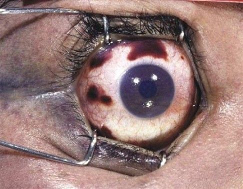 Atrial Septal Defect The heart of this otherwise-healthy 22-year-old Indian woman shows a clear example of an unclosed foramen ovale, which was undiagnosed prior to autopsy. The pulmonary hypertension caused by the left-to-right shunting of blood resulted in acute cor pulmonale and right-heart failure. 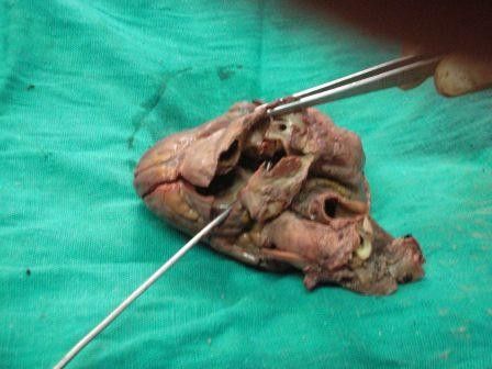 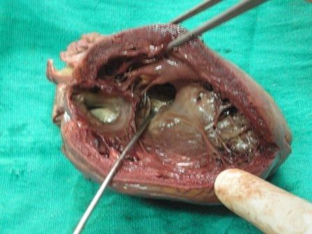 Postmortem blisters are formed 18-24 hours after death due to putrefaction and production of gases which force blood, air and liquid fat into the superficial layers of the skin forming blisters.  Stage IV lung cancer 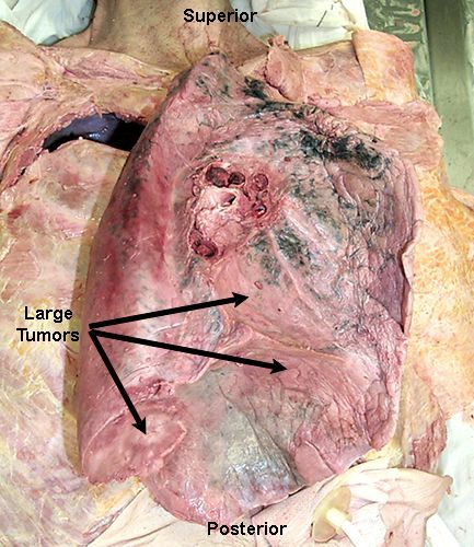 Distant shotgun wound with multiple, evenly distributed discrete defects due to birdshot. 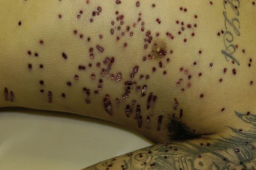 Dissection of the Anatomical Snuffbox Yes, that’s a real area of the body. It’s so called because the triangular deepening on the radial dorsal aspect of the hand was commonly used to place powdered tobacco (snuff) so that it could be sniffed. The anatomical snuffbox is a complex of the three tendons and the artery pictured, with the floor formed by the scaphoid and trapezium bones. 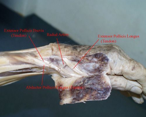 Cerebral hemisphere involvement in a primary CNS lymphoma This is an extremely rare manifestation of B-cell lymphoma. Though still uncommon, AIDS patients (such as the decedent being autopsied) are known to develop this cancer far more often than healthy individuals. Anti-retroviral drugs reduce the incidences of immunodeficiency-related cancers significantly. 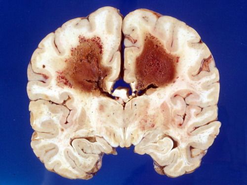 Amputated arm showing effects of gas gangrene Patient was diabetic with poor bowel motility and vascular narrowing. Perforation of bowel mucosa occurred, allowing Clostridium septicum bacteria to infiltrate the bloodstream, causing sepsis and localized gas gangrene, beginning near elbow and extending outwards. 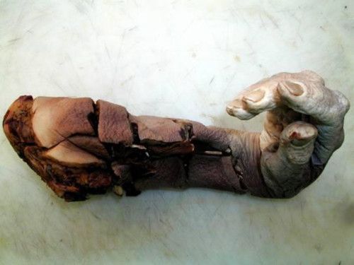 Wet mount example of degloved tissue from decomposing cadaver 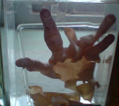 Struck by a car, note the typical impacts to the grill, hood, and windshield (left) and the patterned injuries on the pedestrian (right). 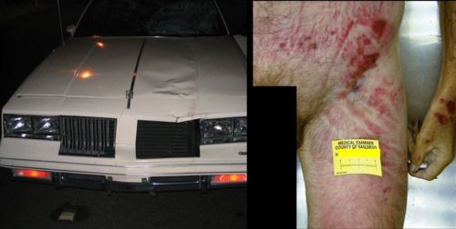 Stellate hard contact wound of the head. Note the abundant soot in the wound depths. 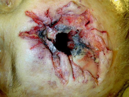 Organs converted to adipocere Though waxy and of different tissue composition, internal organs converted to adipocere can resist putrefaction for many, many years. Adipocere also preserves wounded fatty tissues, allowing for determination of violent deaths involving tissues that would normally decompose (such as a stab wound to the stomach that did not hit any bones). 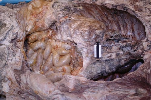 Postmortem artifact of consumption by scavenger on cadaver leg. Vulture activity on leg of deceased woman recovered from forest. Underlying tendon exposed, and much musculature is absent. Irregular scraping yellow defects around the deeper wound characteristic of beak usage. Smaller animals typically leave scalloped edges, and larger animals more jagged edges. The scraping marks and intact tendon are the most telling clues on this artifact. 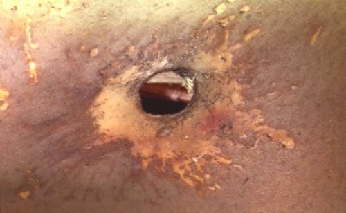 Cadaver late in “bloat” stage of decomposition. You can see advanced skin slippage and de-gloving occurring here. The body will soon begin to deflate, and tissues will liquefy and seep into the surrounding earth 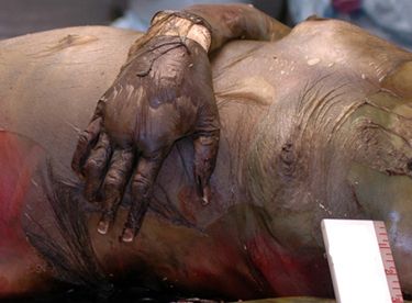 Knife blade inside body Knife broke off at hilt when victim was stabbed. From the outside, it’s difficult to see the mechanism of the intensive internal damage that this victim succumbed to, but an x-ray shows how large of a knife it really was. 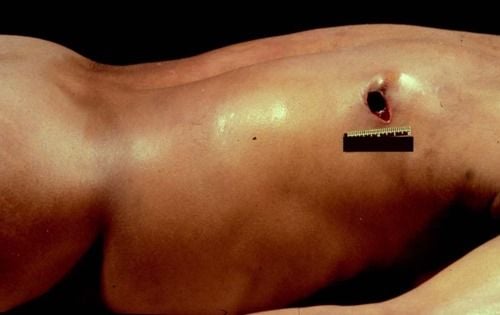 Postmortem consumption of cadaver by alligators Large animals like sharks and alligators may consume chunks of tissue after a person has died, and leave sharp injuries in soft tissue and bone. This can make antemortem trauma (likely meaning that the death was caused by animal attack) and postmortem artifacts difficult to differentiate, making cause of death an uncertainty when nothing else can be found. 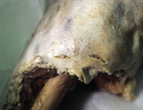 Fungal growth on a previously embalmed and subsequently exhumed body. 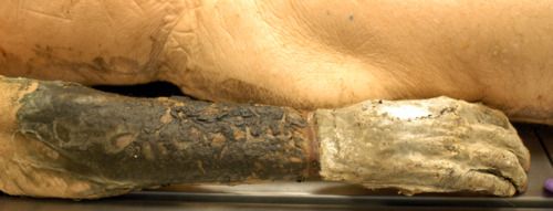 Cadaver Charred in Car Fire Despite loss of the outer layers of the body (including the outer arteries and veins), it’s generally still possible to do all of the internal autopsy, as well as collect toxicology samples. Vitreous fluid (from the eyes) is the most likely to be destroyed or too contaminated to test, but was still found to be available over 40% of the time in Eastern US autopsies performed after fire exposure (where exposure caused 75%-100% of the body to be charred past the dermis). Blood can be obtained from central and femoral areas, rather than peripheral veins. 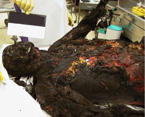 Mummified Homicide Victim Victim remained in secure bedroom in August in Southeastern United States for just under two weeks, with no air conditioner. Though heat often complicates homicide investigations because of increases in insect activity, and because humidity can speed up autolysis and bacterial decomposition, dry heat (like that in Egypt) can actually result in natural mummification. Her incised wounds were so well-preserved by the mummification that they were able to play a key role in prosecution of the perpetrator. 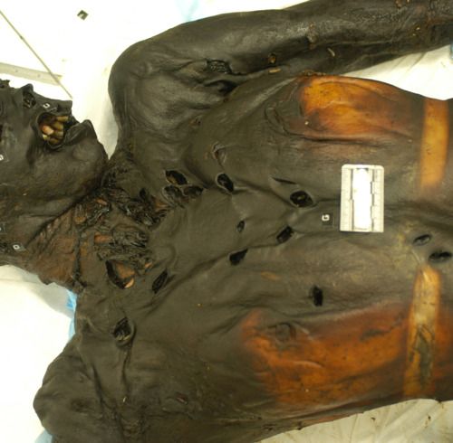 Postmortem gnaw marks on a long bone. You can see the teeth-marks along the ends of the bone, and the lack of symmetrical edging. While asymmetrical edging can occur when a bone is broken (generally very difficult for a human to do unassisted), the ends break in a splintering manner. 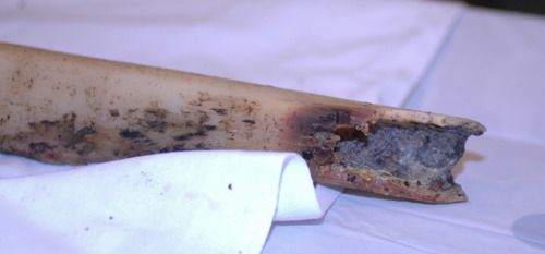 Chop injuries inflicted by boat propeller Cause of death was hypovolemic shock (shock caused by loss of blood volume) on transport to hospital. Decedent had blood alcohol content of 0.12 and was swimming near and below idling boat during river party. Boat operator did not observe him prior to initiating propeller action. Boat operator had BAC of 0.02 (significantly below legal limit to operate moving vehicle). No charges filed. 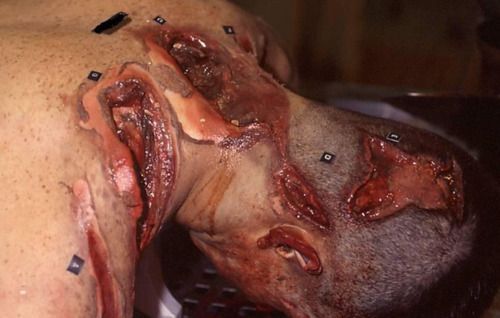 Postmortem entomological activity on left ankle, mimicking ligature marks In cases where the body is not found immediately, forensic entomology is an important subject for a medical examiner to at least know the basics of. The postmortem changes on the left ankle of this victim could be misinterpreted as ligature marks, if the ME didn’t know the telltale signs of insect activity. Entomological activity is also known to mimic blunt-force trauma in certain situations. 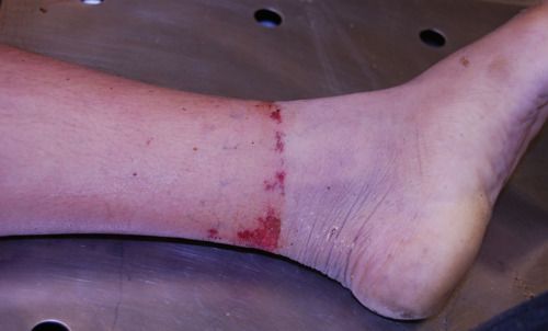 Defensive wounds of hand and fingers When a victim is attacked by someone and attempts to defend themselves, there are often incision wounds (if weapon was sharp) or blunt force trauma and lacerations on the underside of the arms (often used to shield face), the fingers, and the hands. When examining a decedent with defensive wounds, the medical examiner will generally take clippings or swabbings of the undersides of the fingernails of the victim. The presence of the DNA of a suspect can be crucial in proving the suspect’s presence at the attack. 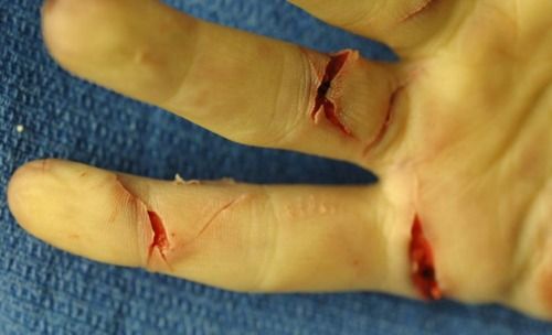 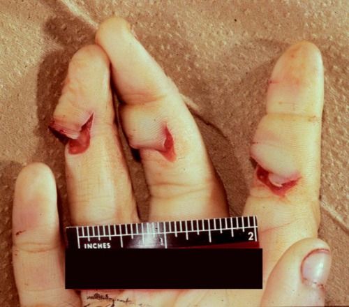 Multiple post-mortem trocar “stab wounds” of heart These types of “wounds” are often caused by the embalming process, and the type and method of embalming (if present) must always be noted when foul play becomes suspected after the decedent has been embalmed, interred, or buried. The cuts made on the side of the neck, in the inguinal region, and the trocar wounds elsewhere, can appear to indicate attacks that did not occur. While experienced medical examiners can fairly easily tell if a body was deceased at the time a wound was inflicted, it can be easy to mistake when less experienced. 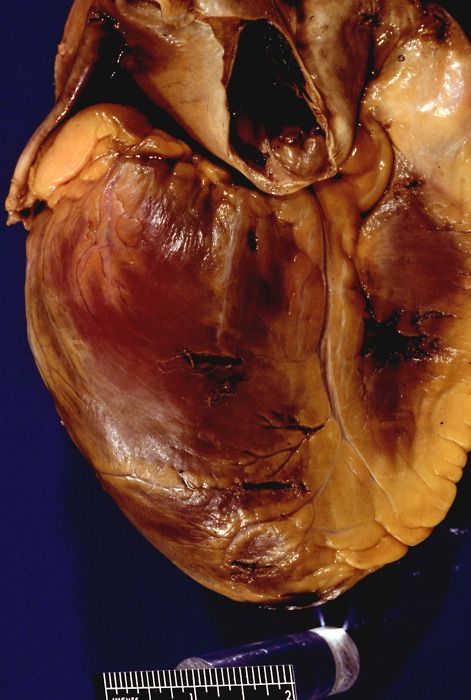 “Chop”-type wound, produced by the claw end of a crowbar. Chopping injuries are interesting, as they display the characteristics of both blunt-force lacerations and sharp trauma. 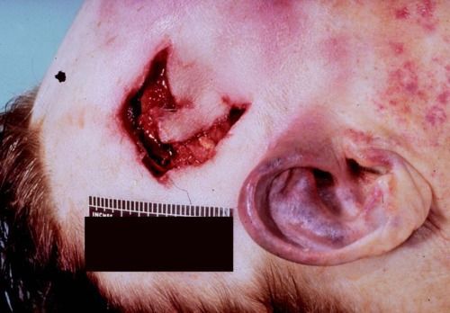 Parts One: http://www.documentingreality.com/fo...s-info-113407/ Part Two: http://www.documentingreality.com/fo...rt-two-114524/ |
|
#4
●
01-13-2013, 04:52 PM
| ||||||||
| My Rank: SERGEANT Poster Rank:958 Join Date: Jun 2012 Posts: 754 Mentioned: 1 Post(s) Quoted: 127 Post(s)
| ||||||||
|
Re: Post-mortem/Dissection Photos With Info Part 3
Really good information! Thank you.  Can't wait to read parts one and two. Thanks again. |
|
#5
●
01-13-2013, 10:19 PM
| ||||||||
 Southern Belle Poster Rank:694 goes off half cocked Join Date: Apr 2012 Posts: 1,249 Mentioned: 0 Post(s) Quoted: 21 Post(s)
| ||||||||
|
Re: Post-mortem/Dissection Photos With Info Part 3
Man, that boat propeller sure did a number on that guy. Note to self: Don't swim near a boat when your drunk  |