|
#1
●
05-16-2022, 09:57 AM
| ||||||||
| ♚ Legacy Gold Member ♚ Poster Rank:908 Join Date: Jul 2020 Posts: 820
Contributions: 10
Mentioned: 13 Post(s) Quoted: 173 Post(s)
| ||||||||
|
Man Commits Suicide with Chainsaw
Australia. This male individual died from lacerated neck injury after committing suicide with a chainsaw. The individual was known to be of high risk of suicide and had recently purchased a chainsaw. There was a large lacerated injury on the anterior aspect of the neck. The edges of the injury appeared relatively smooth. The soft tissues of the neck were completely divided to the anterior aspect of the cervical vertebral bodies. There was obvious division of the bony tissue. The injury measured approximately 15 cm in maximum extent. 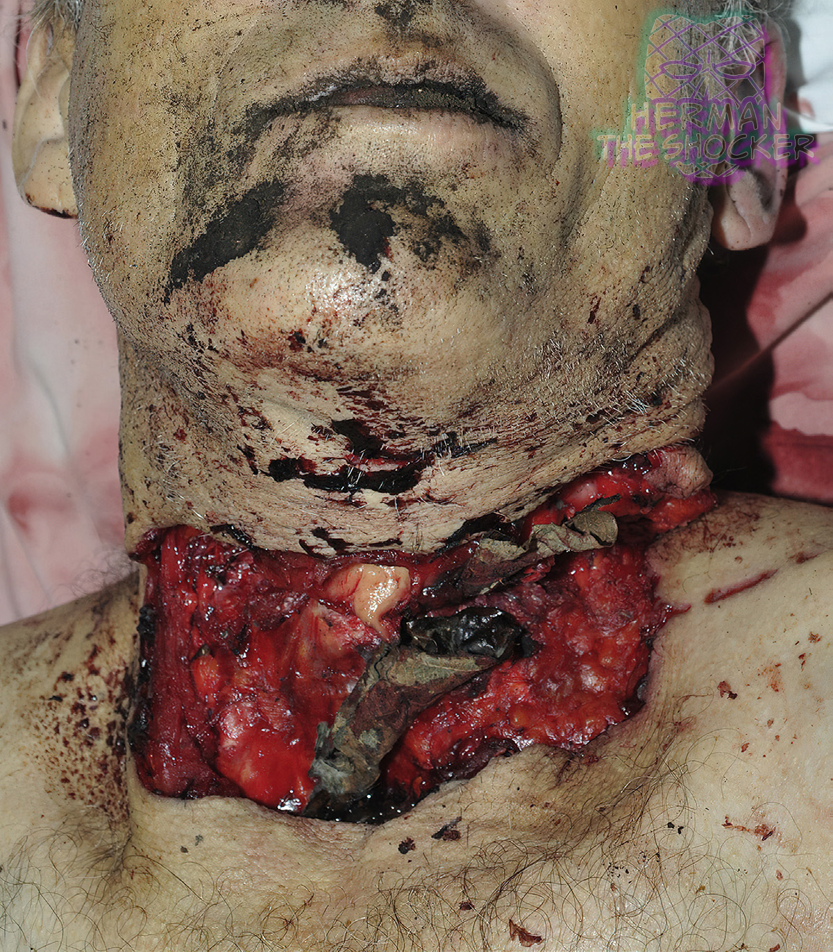 Fig.1 Anterior view of the head and neck showing a large laceration injury to the anterior neck. 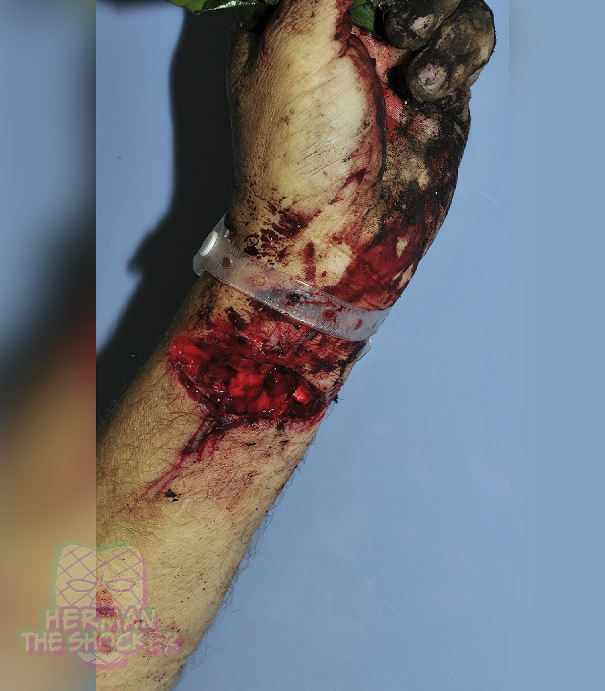 Fig.2 Lateral medial view of the left wrist showing the large laceration. Extending from the left lateral aspect of the injury were discontinuous linear abrasions extending a further 5 cm from the edge of the injury. There were no associated bruises evident. Beneath the chin was a further region of superficial abrasion. There was also a large laceration to the anterior left wrist. The use of a chainsaw is a relatively uncommon means of committing suicide. However, chainsaws have been used in homicide and suicide cases involving adults and children, and typically involve the head or neck. 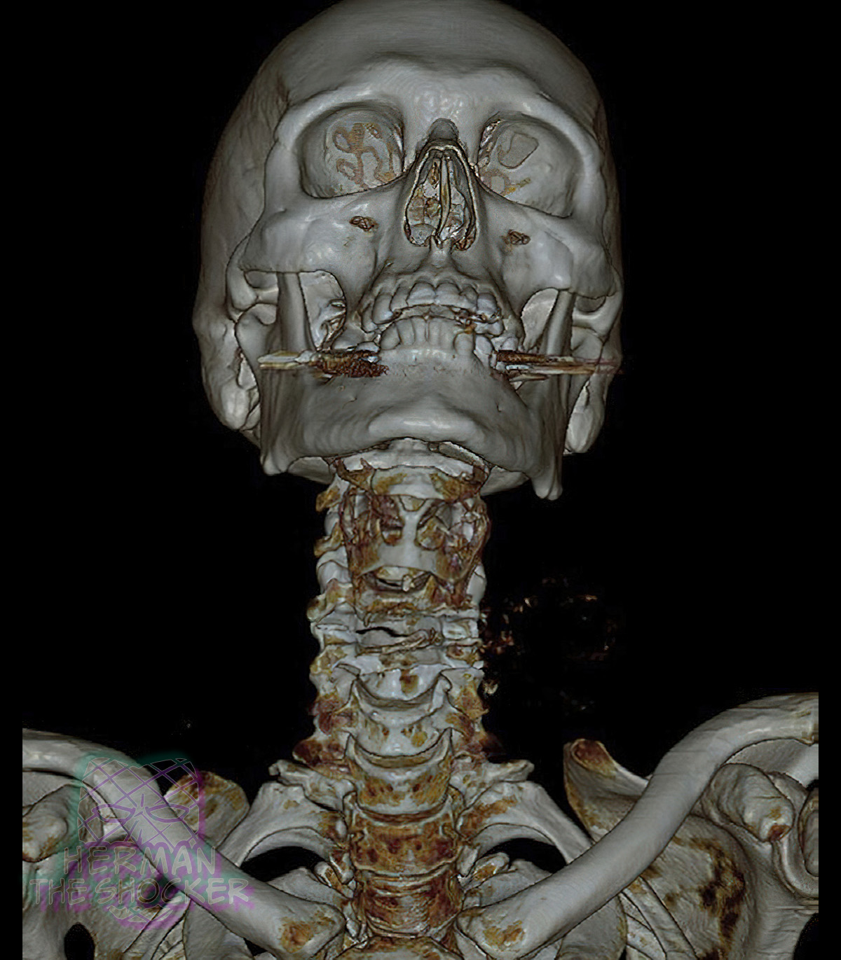 Fig.3 VR image of the anterior skull and vertebrae showing wide dissociation of the larynx and trachea and disruption of two cervical vertebrae. 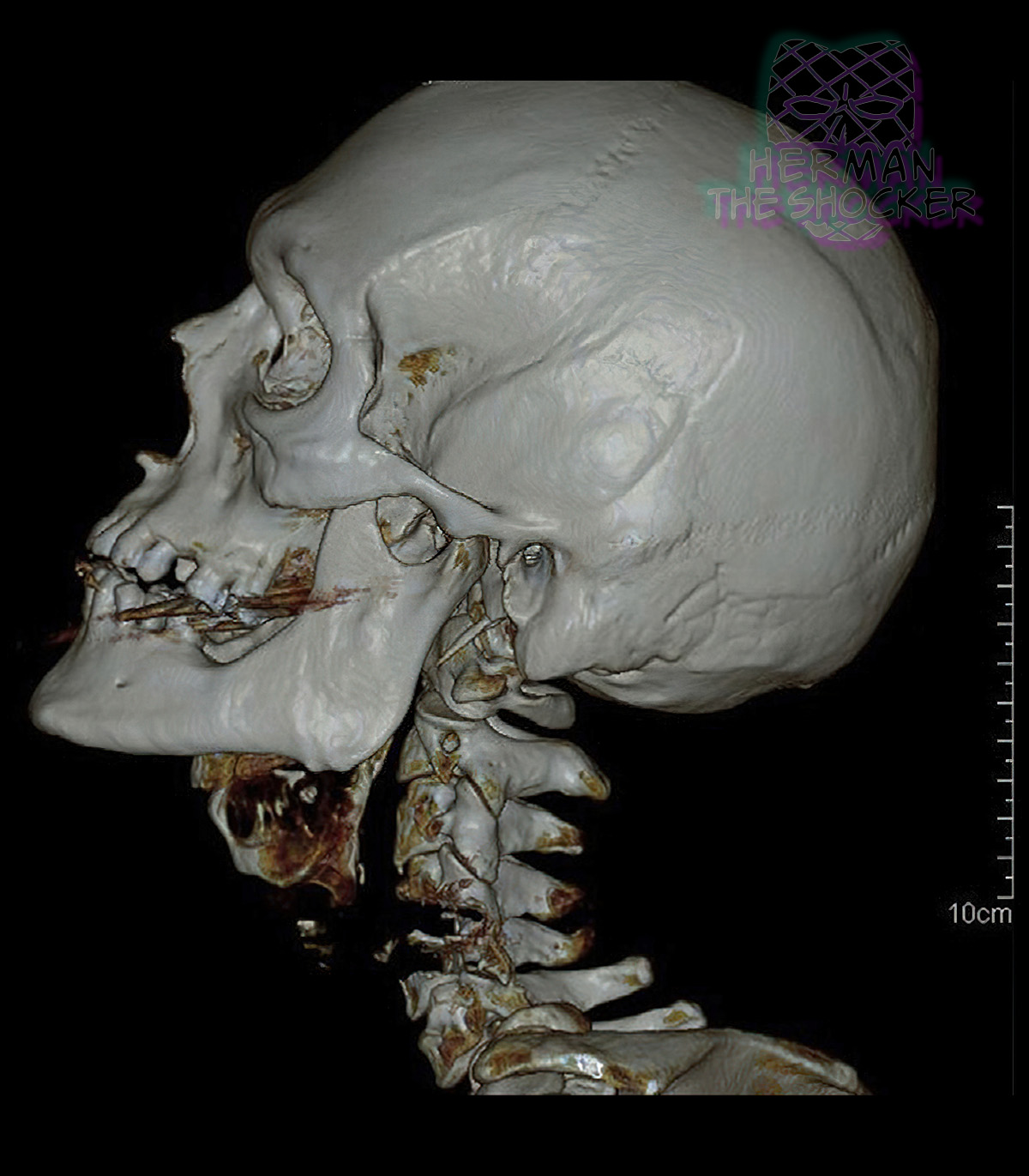 Fig.4 VR image of the left lateral skull and vertebrae showing wide dissociation of the larynx and trachea and disruption of two cervical vertebrae. 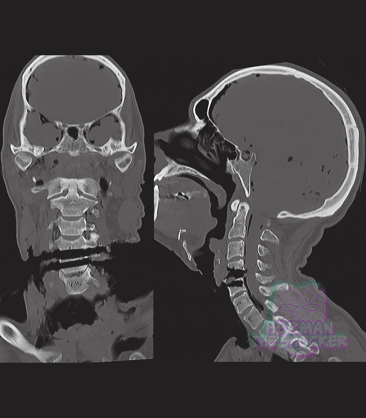 Fig.5 Coronal and sagittal reconstructions of the head and neck showing the parallel linear incised injuries to the 4th and 5th cervical vertebrae. Postmortem CT showed gross disruption to the soft and hard tissues of the anterior neck including wide dissociation of the larynx and trachea, disruption of two of the cervical vertebrae and transection of all major neck blood vessels. The extent of soft tissue disruption was well demonstrated on the volume-rendered surface shaded display, but the depiction of specific bone and soft tissue destruction was best seen on the cross-sectional imaging. There were two well-defined, parallel, transverse, oblique defects in the bodies of the 4th and 5th cervical vertebrae and adjacent soft tissues (measuring ∼6 and 8 mm in width), which extended into the spinal canal; the most superior entered the cervical spinal cord. - This post is for educational purposes only and is nonprofit. Under Section 107 of the US Copyright Act of 1976; Allowance is made for "Fair Use" for purposes such as criticism, comment, news reporting, teaching, scholarship, and research. OP is not a medical expert. No copyright infringement intended. This post does not encourage or glorify violence/harassment. Images might have been upscaled and enhanced. Text might have been shortened and simplified/reorganized for online view.
__________________ ⭐️ hermantheshocker.com ⭐️ |
|
#3
●
05-16-2022, 10:23 AM
| ||||||||
| My Rank: FIRST SERGEANT Poster Rank:394 Join Date: Apr 2011 Posts: 3,054 Mentioned: 2 Post(s) Quoted: 276 Post(s)
| ||||||||
|
Re: Man Commits Suicide with Chainsaw
Definitely not the method for the faint of heart |
|
#5
●
05-16-2022, 12:24 PM
| ||||||||
| ♚ Legacy Gold Member ♚ Poster Rank:445 I'll hoist you upon mine own petard. Join Date: Jul 2010 Posts: 2,552 Mentioned: 5 Post(s) Quoted: 1198 Post(s)
| ||||||||
|
Re: Man Commits Suicide with Chainsaw
When the pain of living outweighs the pain of dying, no method is too extreme.
|
|
#7
●
05-16-2022, 01:33 PM
| ||||||||
| ♚ Legacy Gold Member ♚ Poster Rank:127 Happy Happy Happy Join Date: Dec 2009 Posts: 12,631 Mentioned: 21 Post(s) Quoted: 4171 Post(s)
| ||||||||
|
Re: Man Commits Suicide with Chainsaw
Five out of five corns as always Mr.shocker |
|
#9
●
05-16-2022, 08:24 PM
| ||||||||
| ★ Legacy Member ★ Poster Rank:248 So many choices now Join Date: Jul 2015 Posts: 5,548 Mentioned: 15 Post(s) Quoted: 2120 Post(s)
| ||||||||
|
Re: Man Commits Suicide with Chainsaw
Every CT scan you post could be an album cover, Herm. If I had a band, and this was 1985, we'd both be record-store famous. |


