|
#1
●
07-01-2012, 08:05 AM
| ||||||||
 ★ ********* ★ Poster Rank:251 Male Join Date: Jul 2009 Posts: 5,564
Contributions: 368
Mentioned: 66 Post(s) Quoted: 932 Post(s)
| ||||||||
|
Identification of Human Remains
An important duty of coroners, medical examiners, and forensic pathologists involve s the identification (ID) of deceased individuals. While this process does not usually present many difficulties, the consequences of misidentification can be significant and long lasting. The most common mistake involves the inadvertent misidentification of two bodies as each other. This type of error is most likely to occur in cases of multiple deaths in a single incident, particularly if the deceased individuals are of the same gender and race, with similar ages and sizes. The possibility of such errors is also increased in busy offices where there are many bodies entering and exiting the facility on a daily basis, particularly if there are not well-established identification policies and procedures in place. In many cases investigated by medicolegal death teams, initial inquiries will produce a preliminary or tentative identification. For example, important circumstantial evidence, such as a driver’s license found in the decedent’s possession, may point toward the identity of the deceased. However, a definitive positive identification is required. This can be established by a variety of means with the most common method being visual identification by family members. Although this is most often utilized, it must be recognized that it is not a scientific method, and major mistakes have been known to occur. In addition, in a number of cases visual identification is not possible. This most often occurs when there has been facial disfigurement from trauma, fires, or putrefaction. The most reliable methods of identification involve a more scientific approach where a unique feature of the unidentified decedent can be compared to a likely match. This requires that the features of both are compatible, with no significant features that do not match. Examples of scientific methods of identification include fingerprint comparison, dental comparison and x-ray comparisons, and DNA typing. For each of these methods (except for DNA typing) an antemortem (prior to death) record must exist and be available for a successful comparison to be made. If identity cannot be established by any of the above processes, then other features may be useful in helping to make a positive identification. Examples of other features that can be used include the location and appearance of various birthmarks, scars, implanted medical devices, or tattoos. These may all have features that make them unique to a specific individual, for example, a particular tattoo with names and dates of family members. Finally, the circumstances of the death may provide useful information, for example, a single body in a burnt out house owned by an individual who has not been seen since the fire. Although this is the least certain method of positive identification, it is sometimes all that is available. In a minority of cases a positive identification will not be able to be made. This situation is more common in large metropolitan areas among transient or homeless individuals when no one comes forward to identify the decedent despite the body being visually recognizable. This problem is compounded if bodies are badly decomposed or skeletonized, a situation that occurs in rural areas. When the body is not able to be identified, forensic pathologists will perform the full range of investigations so that if a possible identity is subsequently suggested then all of the necessary information and material for comparison will be available. Such material includes total body photographs, fingerprints, total body x-rays, dental charts and x-rays, and DNA samples (blood, hair, bone, and teeth). Forensic anthropologic assessment of skeletonized remains can also provide useful estimates of the age, race, gender, and stature. On occasion, a forensic artist may attempt a clay facial reconstruction over an unidentified skull, or a computer image of the face may be created, with publishing of such images to the public to find anyone who may recognize the person. 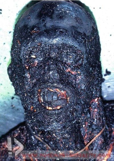 Fig. 1 Bodies recovered from fires may be burnt beyond recognition 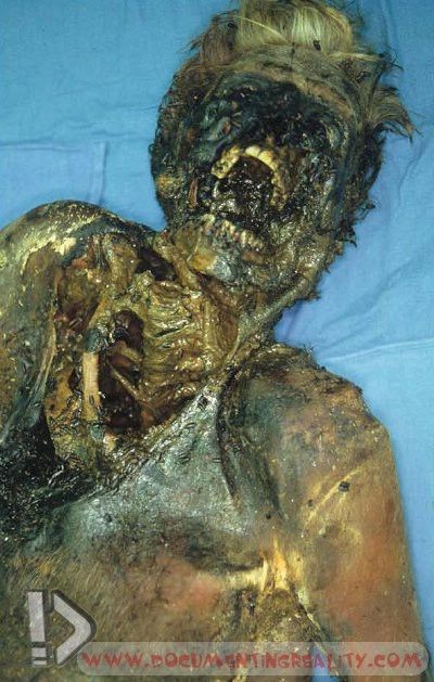 Fig. 2 Badly decomposed bodies are typically not visually identifiable 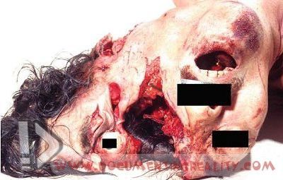 Fig. 3 Depending on the injury type, severity, and location, persons who are victims of severe trauma may not be visually identifiable. This was the victim of a motor vehicle collision 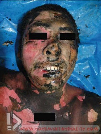 Fig. 4 Even if a fire victim has not been totally charred, they may be burnt beyond recognition, and so a scientific method of identification should be sought 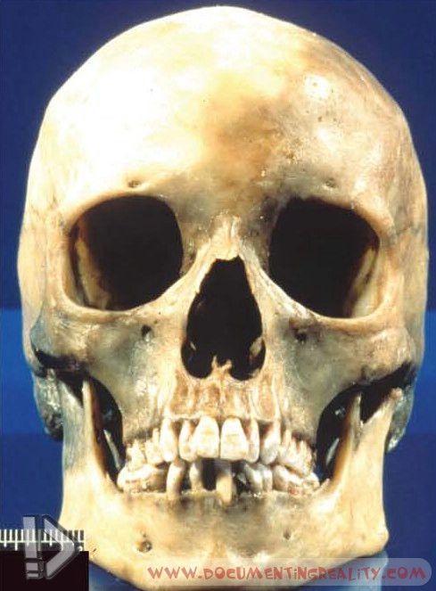 Fig. 5 For obvious reasons, skeletal remains cannot typically be identified visually as there has been complete loss of soft tissue features that determined antemortem morphology 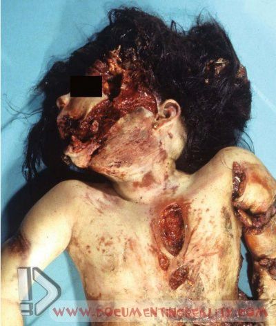 Fig. 6 Another example of severe trauma resulting in facial distortion such that visual identification was impossible Identification of Human Remains 9 203 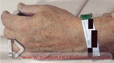 Fig. 7 Hospital identification bands are often considered verification of positive identity 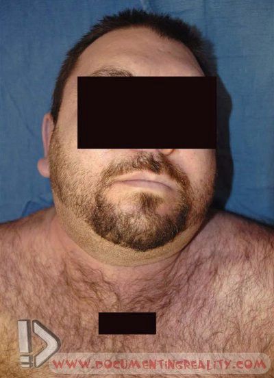 Fig. 8 An ‘‘identification photograph’’ may be shown to a family member to positively establish the identity of a decedent 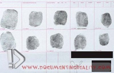 Fig. 9 Many offices routinely take fingerprints on all bodies. This is an example of a ten-print fingerprint card 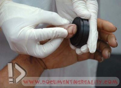 Fig. 10 Ink being applied to a decedent’s finger at autopsy Identification of Human Remains 9 205 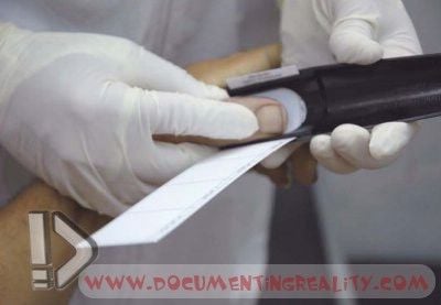 Fig. 11 A fingerprint being taken from a decedent at autopsy 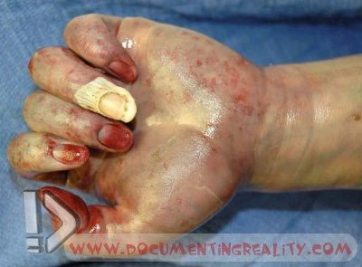 Fig. 12 In certain decomposition cases, all or a majority of the skin of the hands and fingers may slip off. This image shows a hand without most of the skin 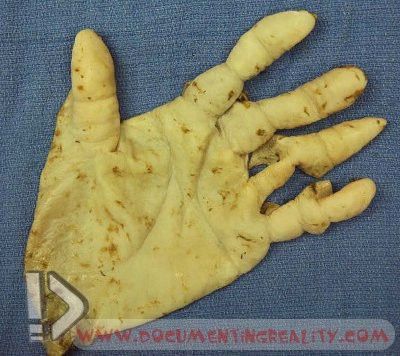 Fig. 13 The ‘‘degloved’’ portion of skin that slipped off the hand shown in >Fig. 12 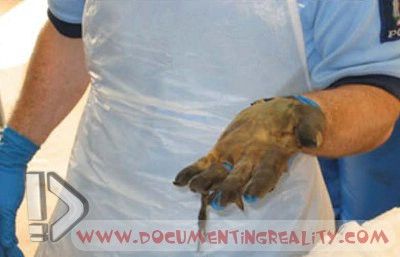 Fig. 14 Skin of the fingers and hand being applied as a ‘‘glove’’ 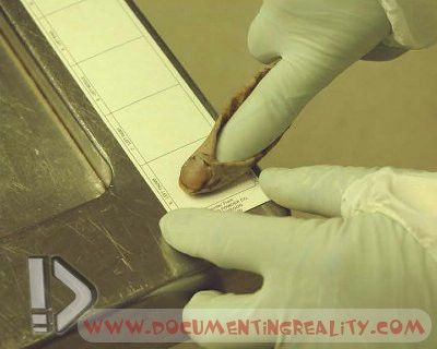 Fig. 15 Using a gloved hand, the pathologist or assistant can place his/her own finger inside the decedent’s finger skin and produce a fingerprint that can be used for identification purposes 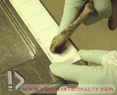 Fig. 16 A fingerprint being produced via a degloved finger 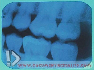 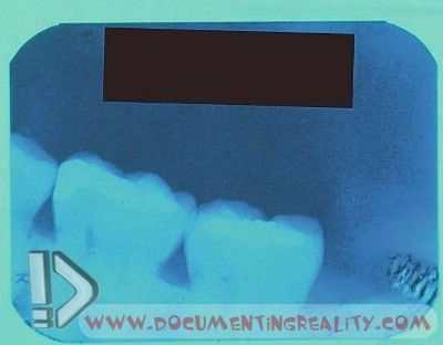 Fig. 17 a + b Above: An antemortem (before death) dental x-ray, received from the decedent’s dentist for comparison purposes. Below: A postmortem dental x-ray performed at autopsy. Comparison of this x-ray to the antemortem x-ray allowed positive identification to be made in this case. Note the unique shapes of the fillings, which appear as radiopaque (white) areas within the teeth 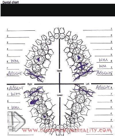 Fig. 18 Besides x-rays, dental charts can be used to make positive identification. This is an example of a dental diagram created at autopsy, for comparison with antemortem records  Fig. 19 A pattern of marked wearing of the teeth with no dental caries or fillings are characteristic features of tribal Aboriginal Australian remains excavated from pre-European contact graves 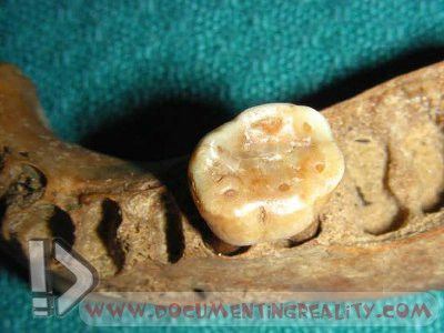 Fig. 20 A closer view of a markedly worn molar tooth from an archaic aboriginal burial in South Australia 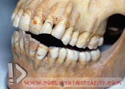 Fig. 21 Notching of the front teeth may be a marker of a long-term pipe smoker 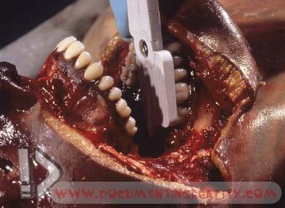 Fig. 22 When a body is not viewable for funeral purposes, dental charting and radiography can be made much easier by first cutting away the skin and soft tissues of the face, thus exposing the underlying teeth. In the example shown, a dental x-ray is being performed 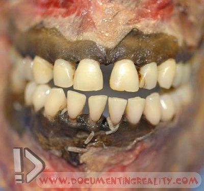 Fig. 23 Another example of a case in which the soft tissues of the face have been removed in order to better visualize the teeth 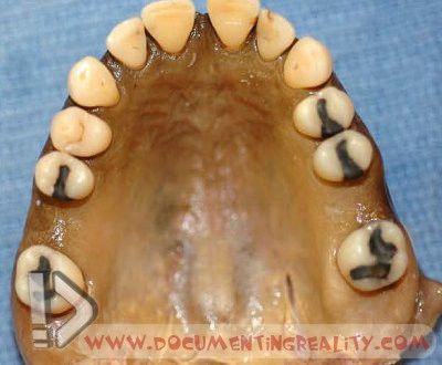 Fig. 24 A maxilla (upper jaw) that has been cut away from the rest of the skull in order to better visualize, and more easily x-ray, the teeth. It is vital that the maxilla is clearly labeled once it has been removed from the body. Loss of identifying labels created significant problems with some of the body identifications during the South-East Asian tsunami 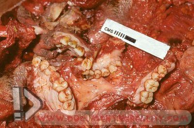 Fig. 25 Despite extensive traumatic disruption, as in this case from a vehicle crash, dental identification may still be possible if fragments of teeth can be retrieved 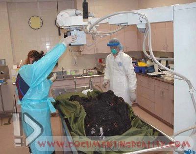 Fig. 26 An x-ray being performed on a badly burned body 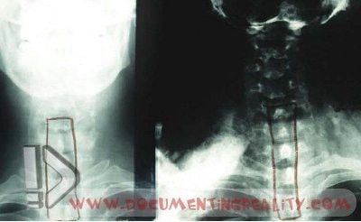 Fig. 27 Left: A close-up view of an antemortem (before death) neck x-ray, received from a hospital for comparison purposes. Right: A close-up view of a postmortem neck x-ray corresponding to the antemortem film. Note that the unique shapes of the spinous processes of the vertebral column in each x-ray are identical, allowing for positive identification 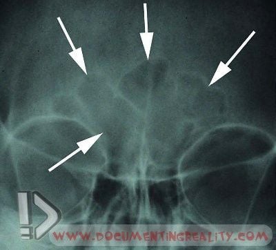 Fig. 28 A skull x-ray, showing the unique shape of the frontal sinuses, which represent radiolucent (darker) air pockets within the skull of the forehead (arrows) 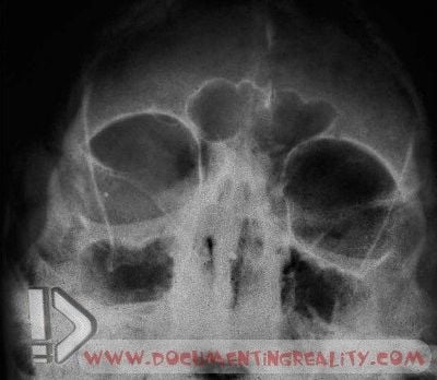 Fig. 29 Another example of an x-ray of the frontal sinuses revealing a unique shape that can be linked to a particular individual 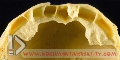 Fig. 30 The top part of the skull that has been cleaned to show the typical air spaces that may be present in the frontal sinuses enabling individual identification to be made 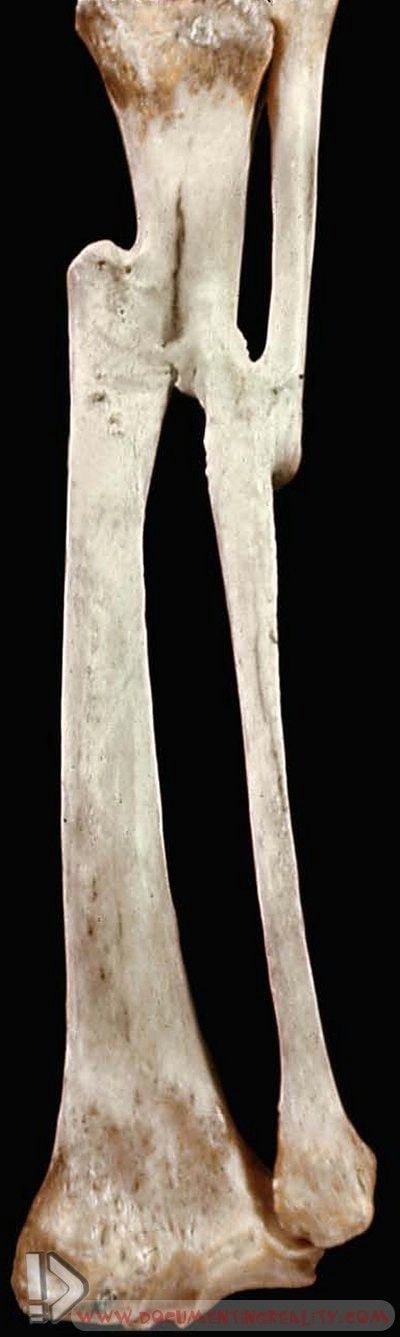 Fig. 31 The site of previous fractures with healing may be useful in identification. In this case a poorly healed fracture of the tibia and fibula of the lower leg would have resulted in shortening of the leg and a history of walking with a limp 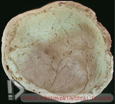 Fig. 32 Sometimes underlying diseases of bone may be present, such as Pagets disease where there is prominent thickening of the skull. This may have resulted in the decedent suffering from deafness 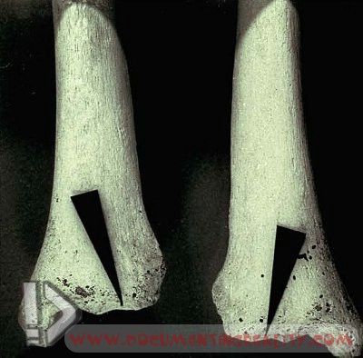 Fig. 33 Skeletal remains from indigenous individuals may show a squatting notch on the lower part of the tibia (arrowheads) 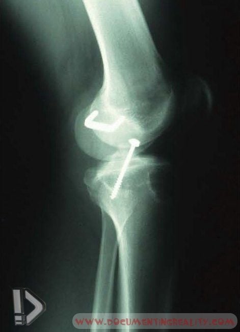 Fig. 34 An x-ray showing orthopedic hardware that was useful in positively identifying a decedent 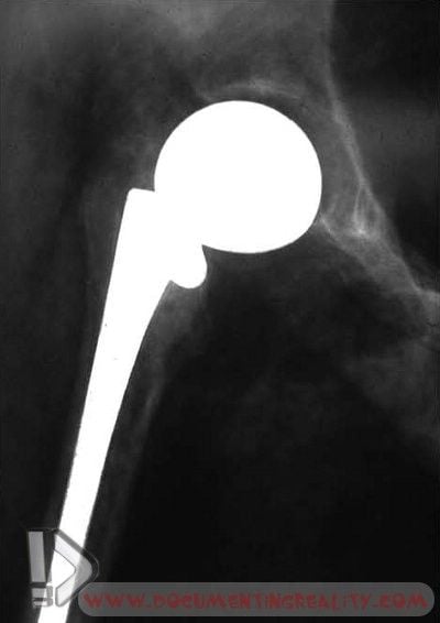 Fig. 35 A postmortem hip x-ray in a badly decomposed woman shows the presence of an artificial hip 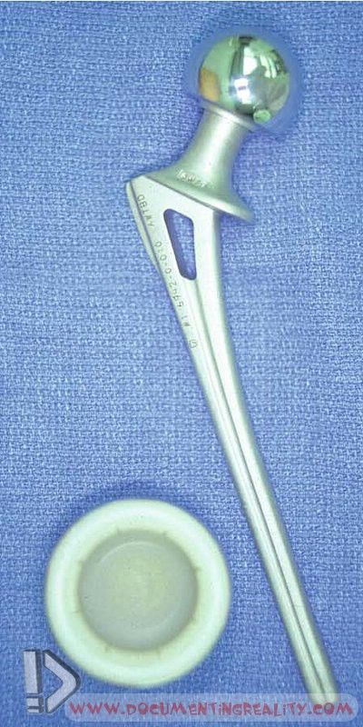 Fig. 36 A photograph of the artificial hip removed at autopsy. Many such implanted devices have unique serial numbers that can be traced, through the manufacturer, to the individual 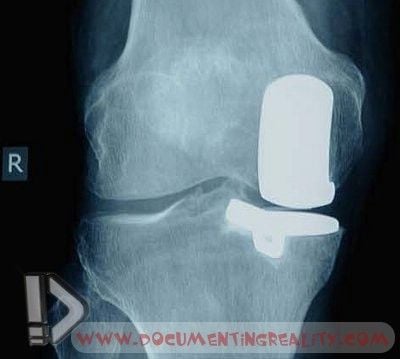 Fig. 37 An x-ray at autopsy may reveal orthopedic appliances such as this knee replacement. Comparison of this x-ray with those taken during life may assist with identification 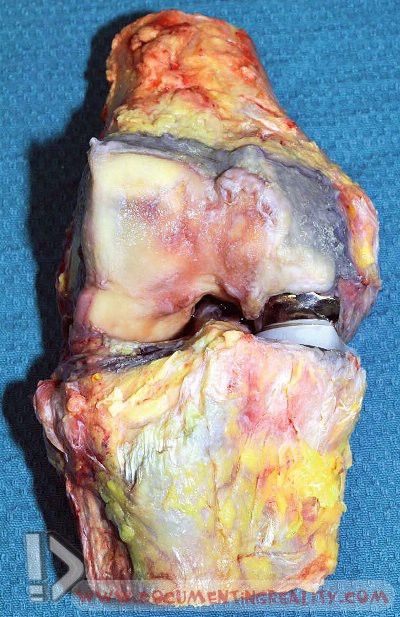 Fig. 38 Dissection of the knee shown in the previous >Fig. 37 reveals the metal and plastic implants 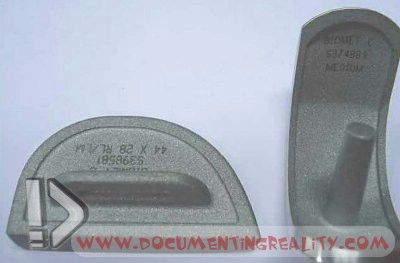 Fig. 39 Once the pieces have been cleaned identifying numbers may be found. Although each piece may not have unique identifying number in every country, the constellation of devices used in one person may be very useful in facilitating identification 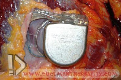 Fig. 40 Implanted medical devices, such as this cardiac defibrillator within the upper left chest, can allow for positive identification to occur at autopsy via unique serial numbers contained on the device and within the medical records 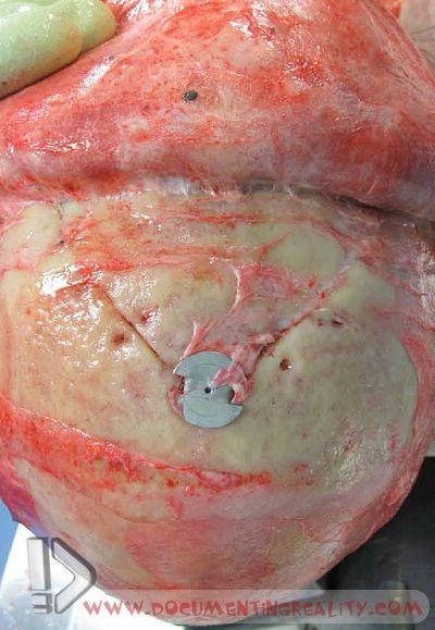 Fig. 41 Sites of previous cranial surgery may be identified on x-ray if there are metal screws or plates. In this case a healed craniotomy incision has been found in a skull at autopsy with a small metal plate 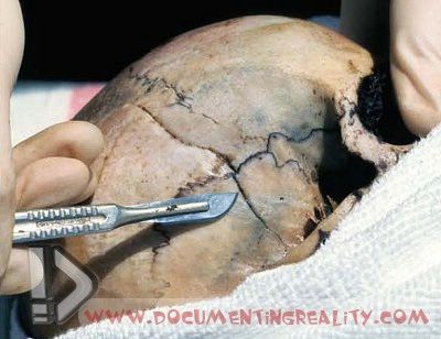 Fig. 42 Another healed craniotomy site identified in skeletonised remains. This is not a fracture 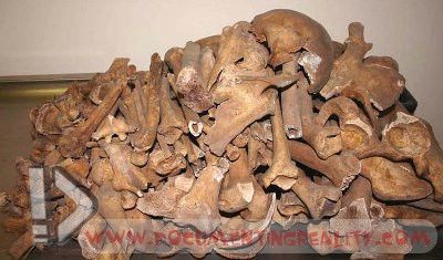 Fig. 43 Large collections of bones may prove to be very difficult to positively identify. These were recently dug up in Berlin, Germany and date from the Battle of Berlin at the end of the Second World War (Photograph provided courtesy of Prof Michael Tsokos, Charite´ University Berlin, Germany) 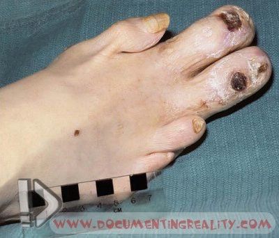 Fig. 44 Certain individuals may have congenital deformities such as these malformed toes that are a unique identifier 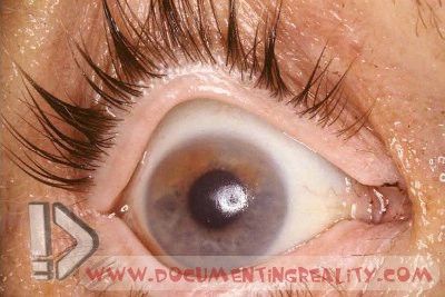 Fig. 45 Some types of injuries may leave subtle markings. The iris in this case has been stained brown in the upper part from a previous perforating metal foreign body 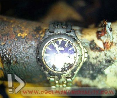 Fig. 46 Jewelry, such as this watch, can be quite valuable in making a positive identification particularly if it is unique or personalized (inscribed with the individual’s name); however, it must be recognized that basing a positive identification solely on such jewelry is not considered scientific 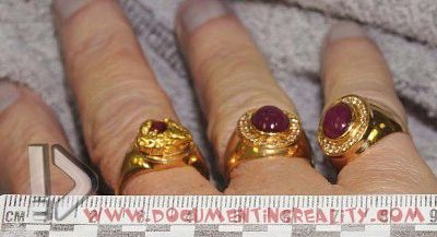 Fig. 47 Fig. 47The rings on these fingers provide useful circumstantial evidence of identity but more scientific methods to establish definitive identification are required  Fig. 48 Piercing sites can be considered further evidence for a positive identification, but should not be the sole criteria utilized as they are not unique 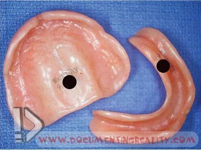 Fig. 49 Dentures, such as those shown in the photo, can be useful in ensuring positive identification. Caution should be exercised, however, since it is not unheard of for elderly individuals, particularly those in extended care facilities and those who are demented, to use other person’s dentures |
|
#2
●
07-01-2012, 08:05 AM
| ||||||||
| ★ ********* ★ Poster Rank:251 Male Join Date: Jul 2009 Posts: 5,564
Contributions: 368
Mentioned: 66 Post(s) Quoted: 932 Post(s)
| ||||||||
|
Re: Identification of Human Remains
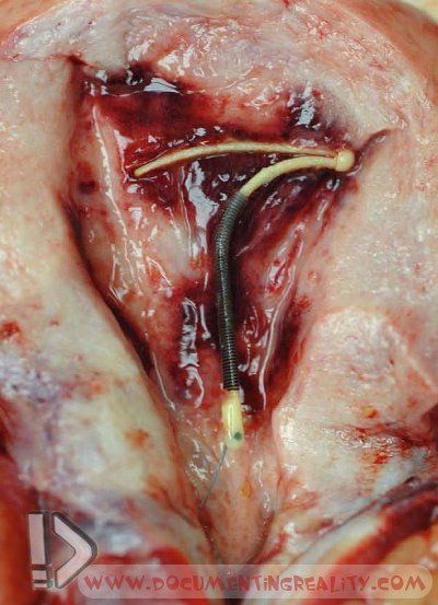 Fig. 50 An intrauterine device found within the uterus at autopsy can provide an additional piece of information useful in making a positive identification 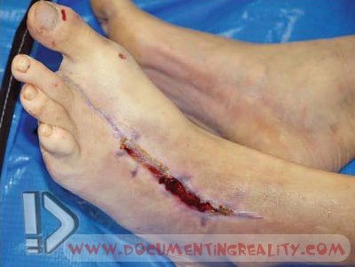 Fig. 51 The history of a recent toe amputation in this decedent was one of several factors which allowed positive identification to be made 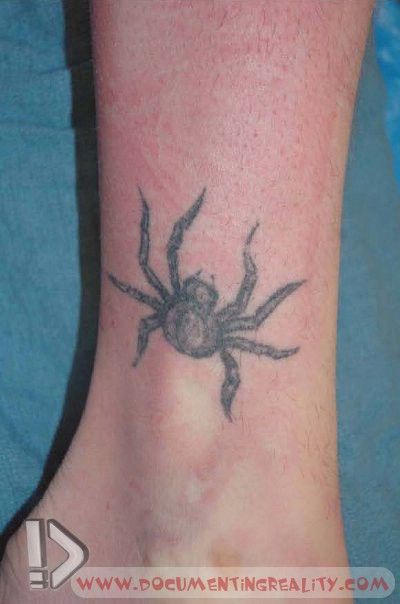 Fig. 52 A tattoo may represent one of several features of a body that allows for positive identification 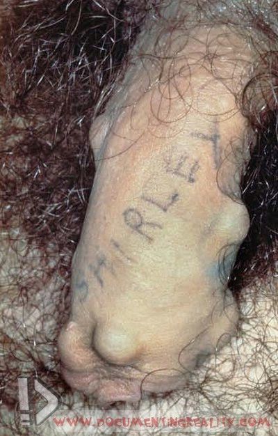 Fig. 53 The implantation of plastic balls under the skin of the penis with the tattoo of a woman’s name may provide unique identifying information  Fig. 54 Penile implants in conjunction with tattoos are also useful in identification  Fig. 55 Tattoos of eyes over the buttocks are an unusual design that may assist with identification 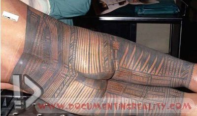 Fig. 56 Tribal tattoos such as the one depicted here from the South Pacific may indicate that the decedent came from a particular cultural or ethnic group 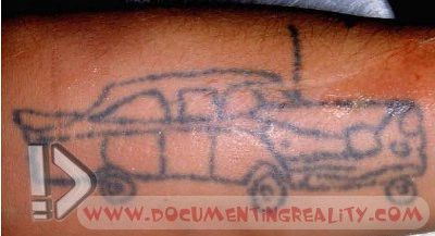 Fig. 57 Tattoos may be extremely rudimentary indicating application at home or in prison. Certain prison groups, such as those from Russia or the former Soviet Union, may have extensive tattooing providing a history of crimes committed, convictions and subsequent incarcerations  Fig. 58 Another primitive tattoo 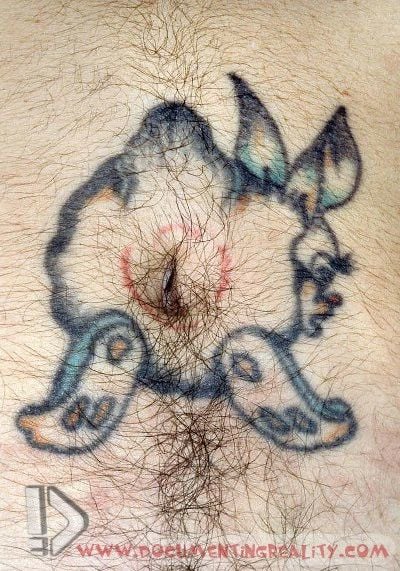 Fig. 59 On occasion, certain body parts such as the umbilicus may be incorporated into a tattoo design  Fig. 60 Modern fine line tattoos may be extremely sophisticated  Fig. 61 Extensive whole body tattoos are found in certain criminal groups such as the yakuza from Japan 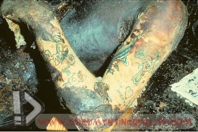 Fig. 62 Tattoos may survive incineration and may also be demonstrated more clearly in an area of burns if the superficial skin layers are removed 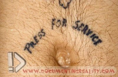 Fig. 63 Humorous tattoos tend to be individualized 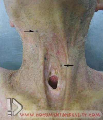 Fig. 64 To assist in positioning radiotherapy beams for the treatment of cancer small dots may be tattooed onto the skin over the tumor. In this case the tracheostomy indicates that there was a cancer of the larynx that was surgically removed. The arrows point to radiotherapy tattoos 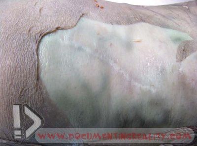 Fig. 65 Identifying scars may be revealed more clearly in decomposed bodies if the surface layer of skin is removed 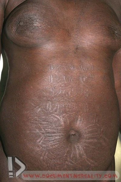 Fig. 66 Scarification in individuals from Africa may be very useful in identifying the specific tribal group that the decedent belonged to. Symmetrical designs can be seen around the umbilicus and upper abdomen 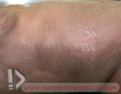 Fig. 67 Tribal scarification of the flank 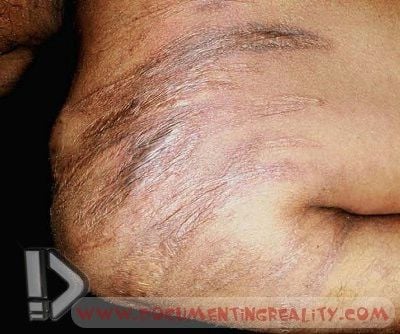 Fig. 68 ‘‘Sorry’’ scars on the shoulder and upper arm of an Australian Aboriginal male that were inflicted as part of traditional mourning ceremonies  Fig. 69 Subincision of the penis is another marking that may be used to identify a traditional Australian Aboriginal male 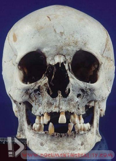 Fig. 70 Occasionally, skeletonized remains are examined at a medioclegal death investigation office, but there is absolutely no indication of the identity of the decedent, either by body exam or scene/ circumstance. After forensic anthropologic examination to characterize the race/ethnicity, gender, and approximate age of the individual, the forensic pathologist may choose to send the skull to a forensic artist, who then attempts to reconstruct the facial features of the decedent (see >Fig. 71) 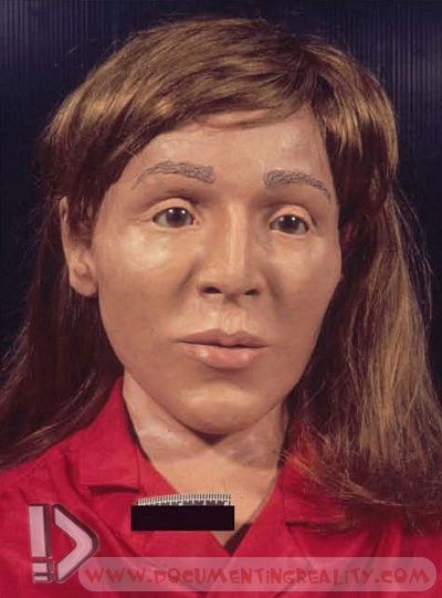 Fig. 71 A forensic artist’s facial reconstruction over the skull shown in >Fig. 70. A photo such as this can be published, with the hope that someone might recognize the person. If someone believes that the decedent might be a particular person, then scientific methods for identity can be undertaken  Fig. 72 In mass disasters all types of identification methods may be utilized. Here, a large number of putrefying bodies have been brought in for identification following the 2004 Boxing Day Tsunami in Thailand Source: The Atlas of Forensic Pathology for Police, Forensic Scientists, Attorneys and Death Investigators (2012)  References Blau S, Hill A, Briggs CA, Cordner SM. Missing persons-missing data: the need to collect antemortem dental records of missing persons. J Forensic Sci. 2006;51:386–9. ByardRW, Simpson E.Characteristic acquired features of indigenous Australians that may be observed in forensic practice. Forensic SciMed Pathol. 2005;1:207–13. Byard RW, James RA, Zuccollo J. Potential confusion arising frommaterials presenting as possible human remains. Am J Forensic Med Pathol. 2001;22:391–4. Byard RW, Simpson E, Both K. The identification of submerged skeletonised remains. Am J Forensic Med Pathol. 2008;29:69–71. Cains G, Byard RW. The forensic and cultural implications of tattooing. In: Tsokos M, editor. Forensic pathology reviews. Totowa: Humana Press; 2008. p. 197–220. Fields R, Molina DK. A novel approach for fingerprinting mummified hands. J Forensic Sci. 2008;53:952–5. HaglundWD, Sperry K. The use of hydrogen peroxide to visualize tattoos obscured by decomposition and mummification. J Forensic Sci. 1993;38:147–50. Hanzlick R, Clark S. The unidentified decedent reporting system – a model national website registry for the unidentified deceased. Am J Forensic Med Pathol. 2008;29:106–13. Hanzlick R, Smith GP. Identification of the unidentified deceased: turnaround times, methods, and demographics in Fulton County, Georgia. Am J Forensic Med Pathol. 2006;27:79–84. Hollmann T, Byard RW, Tsokos M. The processing of human bones found in Berlin, Germany. J Forensic Leg Med. 2008;15:420–5. Murphy WA, Spruill FG, Gantner GE. Radiologic identification of unknown human remains. J Forensic Sci. 1980;25:727–35. Paulozzi LJ, Williams DD, Nolte KB. John and Jane Doe: the epidemiology of unidentified decedents. J Forensic Sci. 2008;53:922–7. Simpson E, Byard RW. Unique characteristics at autopsy that may be useful in identifying human remains. In: Tsokos M, editor. Forensic pathology reviews. Totowa: Human Press; 2008. p. 175–95. Simpson E, James RA, Eitzen D, Byard RW. Role of orthopedic implants and bone morphology in the identification of human remains. J Forensic Sci. 2007;52:442–8. |
|
#5
●
07-01-2012, 09:52 AM
| ||||||||
 ★ Legacy Member ★ Poster Rank:72 male Join Date: Apr 2010 Posts: 21,199
Contributions: 1
Mentioned: 52 Post(s) Quoted: 4957 Post(s)
| ||||||||
|
Re: Identification of Human Remains
thank goodness for teeth, the maggots can't eat them  Great post Gustavo |