|
#1
●
10-07-2021, 08:08 AM
| ||||||||
 ♚ Legacy Gold Member ♚ Poster Rank:908 Join Date: Jul 2020 Posts: 820
Contributions: 10
Mentioned: 13 Post(s) Quoted: 173 Post(s)
| ||||||||
|
2 Cases of Brain Tissue in the External Auditory Canal
In closed-head injuries, the presence of brain tissue in the external auditory canal indicates a hinge fracture (usually including petrous parts of both temporal bones) with dura laceration and separation of the skull base into halves. This phenomenon also demonstrates that the head injury occurred because excessive impulse force or considerable pressure was applied to the head, i.e. the head was com- pressed and/or squeezed. - - - Case1 A 26-year-old male was fatally injured as a helmet-wearing motorcyclist when, after collision, a truck ran over his head and trunk. The left external auditory canal showed the presence of brain tissue (Fig. 2). His head was deformed due to side-to-side compression (Fig. 1). Autopsy findings comprised multiple head fractures (including the petrous part of the temporal bones – hinge fracture), multiple dura and brain lacerations, as well as multiple inner organ injuries and bone fractures. 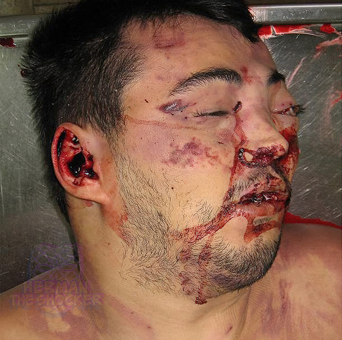 Fig.1 Case1. Head deformation due to side-to-side compression. 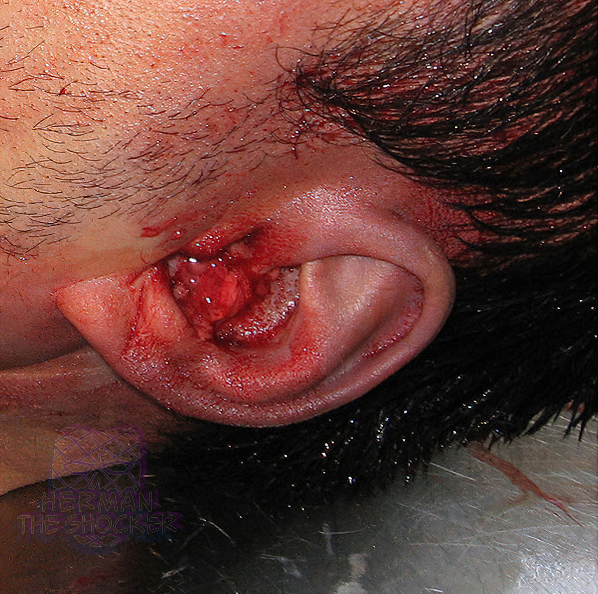 Fig.2 Case1. Brain tissue in the left external auditory canal. 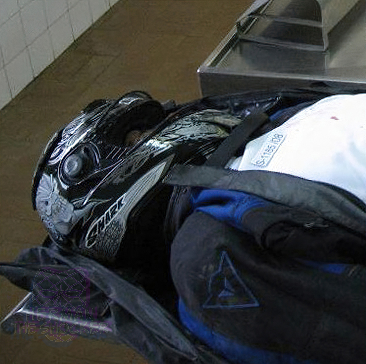 Fig.3 Case1. Fatally injured motorcyclist with his helmet on (photograph taken before autopsy). - - - Case 2 A 33-year-old male committed suicide by jumping from a building about 20 m (65 ft) high, impacting the solid surface feet-first (Figs. 4-5). External examination at the scene of death revealed the presence of blood and tiny brain tissue particles in the left external auditory canal (Fig. 6). Autopsy findings comprised multiple head fractures (including the petrous part of the temporal bones and occipital bone – ring fracture) (Fig. 7), multiple dura and brain lacerations, as well as multiple inner organs injuries and bone fractures.  Fig.4 Case2. Body position after suicidal jump. 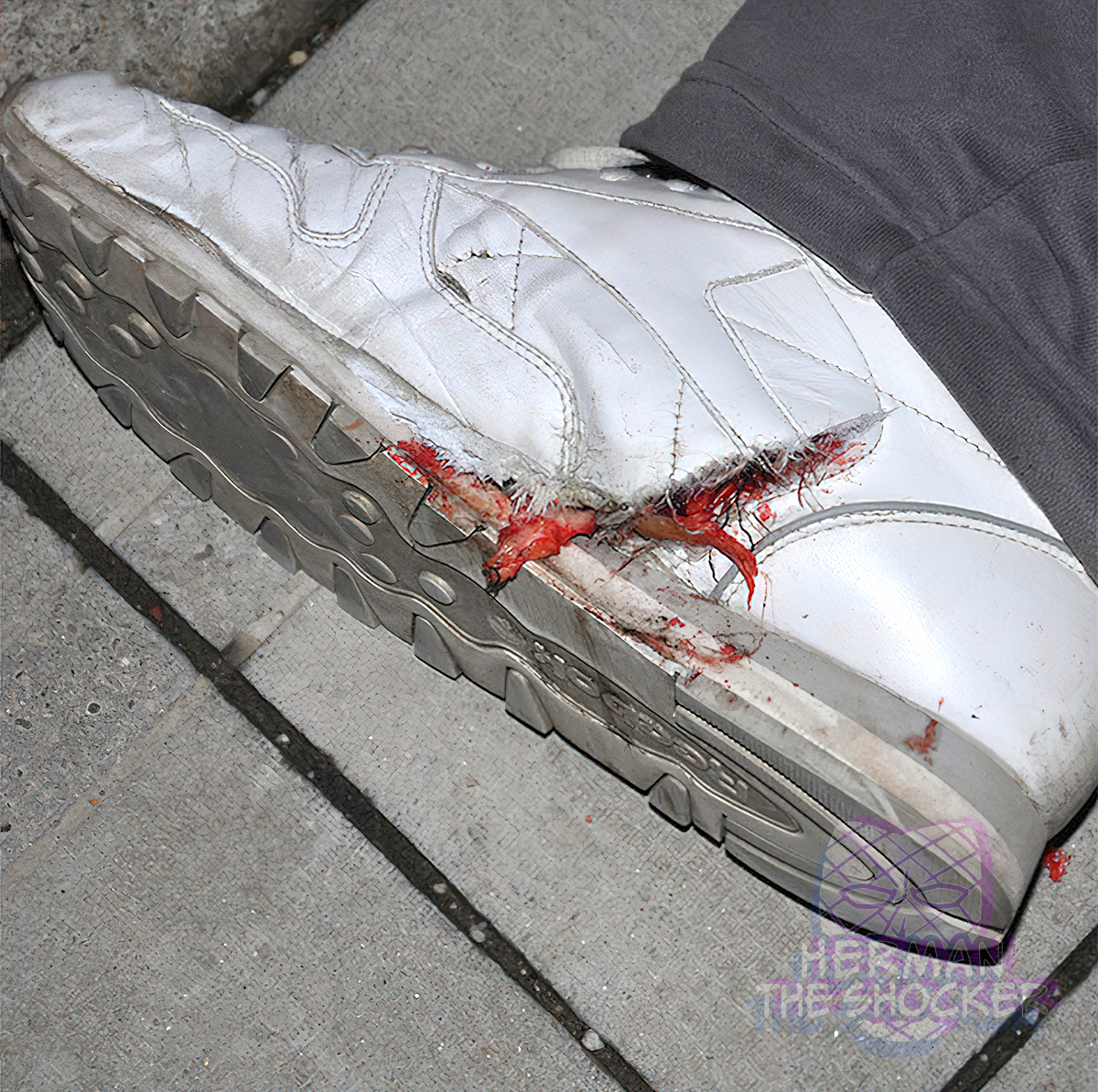 Fig.5 Case2. Damage of the inner side of the shoes with detached soft tissue protruding through it. 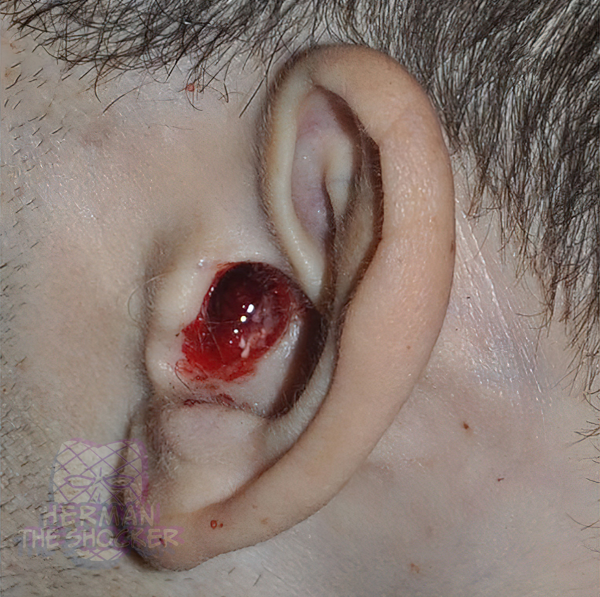 Fig.6 Case2. Brain tissue remains drifting in a small amount of blood in the right external auditory canal. 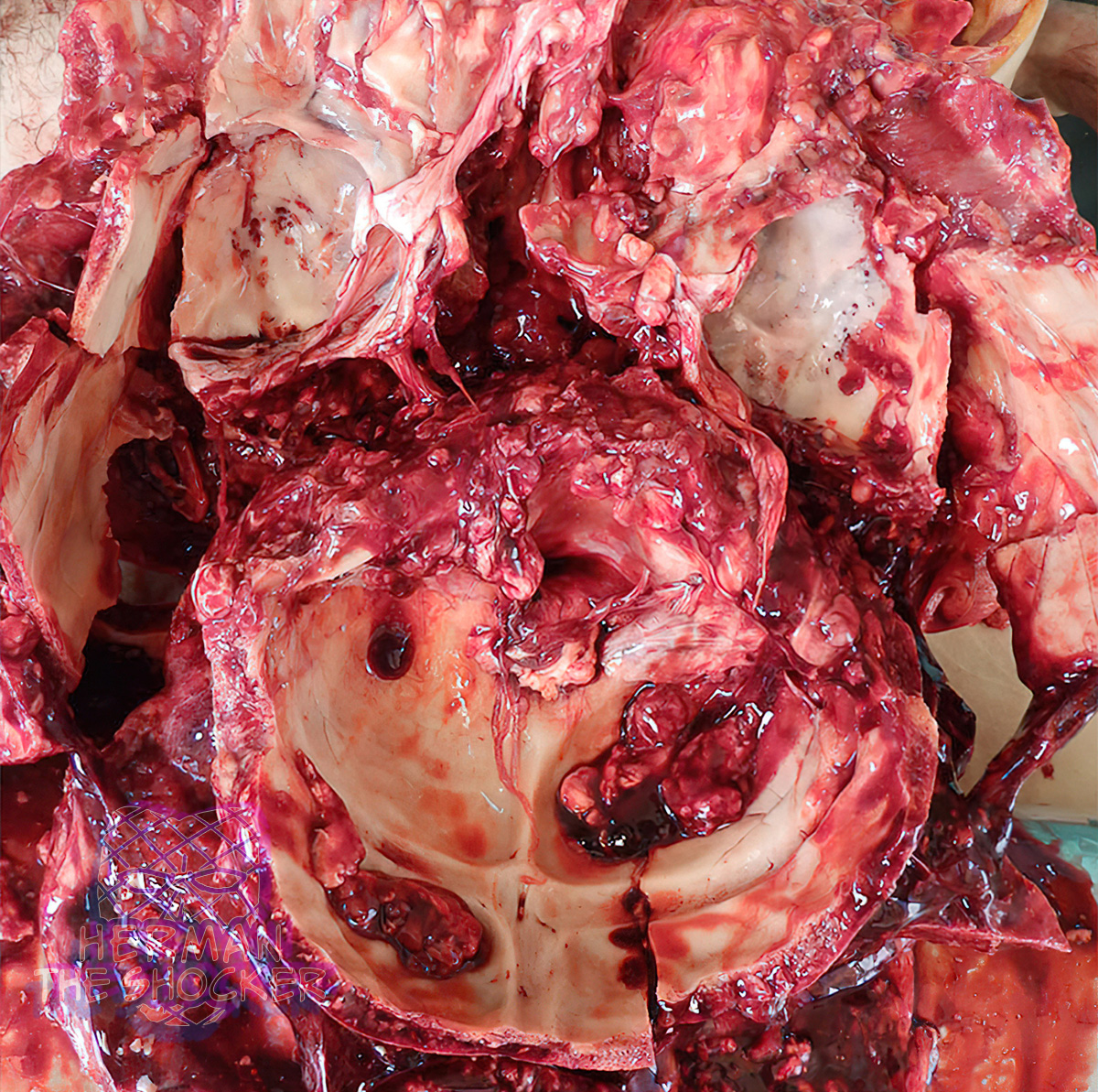 Fig.7 Case2. Ring fracture of the skull base. - - - Bleeding from the ears, nose, or mouth following a closed-head injury suggests a fracture of the base of the skull. Brain tissue could be expelled through these apertures, even in the absence of external evidence of an injury. The presence of brain tissue in the external auditory canal is a relatively uncommon phenomenon, but one that could be expected in side-to-side head compression. In those cases, the force applied to the head is considerable, resulting in excessive pressure and kinetic energy being transferred to the brain tissue. One of the mechanisms of the hinge fracture is also side-to-side head compression. Many basilar skull fractures originate from impacts at the roughly same level as the skull base and the fracture lines follow predictable routes through the skull base. A hinge fracture refers to a fracture that runs from side to side, across the floor of the middle cranial fossa, passing through the pituitary fossa in the midline. This injury is common in road accidents and is sometimes called the “motorcyclist’s fracture”. From the site of applied force, which is usually bitemporal (latero-lateral), fracture lines lead to the separation of the petrous part of the temporal bone from the greater wing of the sphenoid and the squamous temporal bone, splitting the base of the skull into two. Any considerable pressure applied to the head pushes brain tissue equally in all directions, due to its incompressibility. Brain tissue then passes through the lacerated dura into the fractured petrous part of the temporal bones, finally reaching the middle ear cavity. Since this must cause a rupture in the tympanic membrane, the blood and brain tissue ends up in the external auditory canal which is why they can easily be detected at external examination (as seen in this case). The tissue follows the path of least resistance, thus being literally “thrust into the ears” as the result of the high pressure applied on the head. This could be easily demonstrated by using a simple autopsy technique, the so-called” water test “– water poured in an upwardly facing ear result in drainage from the nose and opposite ear, thus demonstrating transcranial passage. The mechanism of injury could be squeezing or compression of the head, either between two solid objects or as the result of being run over by a vehicle. If the pressure to the head is considerable and the body of the sphenoid bone is crushed, the pieces of detached brain tissue can reach the nasopharynx, larynx, and even trachea. In these cases, the presence of brain tissue in the trachea should be considered a purely mechanical phenomenon (the tissue is forcefully pushed), which means that it was not a vital reaction, i.e. brain aspiration. - This post is for educational purposes only and is nonprofit. Under Section 107 of the US Copyright Act of 1976; Allowance is made for "Fair Use" for purposes such as criticism, comment, news reporting, teaching, scholarship, and research. OP is not a medical expert. No copyright infringement intended. This post does not encourage or glorify violence/harassment. Images might have been upscaled and enhanced. Text might have been shortened and simplified/reorganized for online view. |
|
#2
●
10-07-2021, 08:47 AM
| ||||||||
 ♚ Legacy Gold Member ♚ Poster Rank:26 ㅤㅤ真 Join Date: Apr 2021 Posts: 52,944 Mentioned: 372 Post(s) Quoted: 41105 Post(s)
| ||||||||
|
Re: 2 Cases of Brain Tissue in the External Auditory Canal
brutal. 
__________________ ᓚᘏᗢ |
|
#3
●
10-07-2021, 10:12 PM
| ||||||||
 ♚ Legacy Gold Member ♚ Poster Rank:905 Join Date: Oct 2020 Posts: 822 Mentioned: 0 Post(s) Quoted: 119 Post(s)
| ||||||||
|
Re: 2 Cases of Brain Tissue in the External Auditory Canal
That one dude has brains coming out of his nice white tennis shoes too. Imagine that, 2 cases auditory and 1 metatarsal...amazing! Plus, he's the only guy I've ever seen that looks shorter lying down. |
|
#4
●
10-07-2021, 10:24 PM
| ||||||||
 ★ Legacy Member ★ Poster Rank:248 So many choices now Join Date: Jul 2015 Posts: 5,548 Mentioned: 15 Post(s) Quoted: 2120 Post(s)
| ||||||||
|
Re: 2 Cases of Brain Tissue in the External Auditory Canal
Fig 3, Case 1 - Non-helmet types will argue the point that a helmet didn't save this guy's life, so why wear one?  |
|
#5
●
10-07-2021, 11:39 PM
| ||||||||
 My Rank: MASTER SERGEANT Poster Rank:481 Female Join Date: Jul 2021 Posts: 2,168 Mentioned: 60 Post(s) Quoted: 2078 Post(s)
| ||||||||
|
Re: 2 Cases of Brain Tissue in the External Auditory Canal
That's an awesome helmet. It almost makes wearing a helmet look cool.
|
|
#6
●
10-08-2021, 07:52 AM
| ||||||||
| So Fucking Banned Poster Rank:389 Join Date: Nov 2016 Posts: 3,080 Mentioned: 9 Post(s) Quoted: 1583 Post(s)
| ||||||||
|
Re: 2 Cases of Brain Tissue in the External Auditory Canal
I'm going to move to the country and never leave it. I don't want die in a fucking parking garage. I want to die while riding in the vast, natural, empty expanse - on Planet Mars!
|
|
#7
●
10-08-2021, 01:35 PM
| ||||||||
 My Rank: STAFF SERGEANT Poster Rank:805 I Am Dic-N-Bawss Join Date: Aug 2012 Posts: 967 Mentioned: 0 Post(s) Quoted: 176 Post(s)
| ||||||||
|
Re: 2 Cases of Brain Tissue in the External Auditory Canal
I like the thumbnail you got there El Guapo. Is that Hunter "Yeah it's true, I banged my Sister and then my Niece while smoking crack and shooting NyQuil, so what!" Biden? |