|
#1
●
06-06-2025, 03:44 PM
| ||||||||
| My Rank: PRIVATE Poster Rank:33883 Join Date: May 2024 Posts: 1 Mentioned: 0 Post(s) Quoted: 0 Post(s)
| ||||||||
|
Systemic Epidermal Nevus Due to FGFR3 Mutation
CASE REPORT Systemic epidermal nevus with involvement of the oral mucosa due to FGFR3 mutation Background Epidermal nevi (EN) are benign hamartomas of the skin arising from the embryonic ectoderm. Depending on the involved components of the epidermis, EN are further divided into organoid and non-organoid (keratinocytic) types [1]. They are usually present at birth or develop during the first years of life, and their incidence is estimated to be 1-3 per 1000 live births [2]. Keratinocytic nevi typically follow the lines of Blaschko. Systemic keratinocytic nevi are characterized by an extensive involvement of large skin areas and may be associated with skeletal, cerebral or ocular abnormalities, resulting in various types of EN syndromes [1,2]. EN represent genetic mosaicism of the skin and activating FGFR3 (Fibroblast Growth Factor Receptor 3) and PIK3CA point mutations have recently been identified in keratinocytic nevi [3-6]. We report a patient with systemic EN associated with a slight scoliosis, who displayed mosaicism of the R248C FGFR3 mutation in epidermis, oral mucosa and blood leukocytes. Case presentation A 17-year old girl was referred with widespread EN (Figure 1 A+B). She was otherwise healthy apart from a tendency to back pain. Her parents recalled the first appearance of the EN when she was 4 months old. The EN initially presented as hyperpigmented linear streaks which gradually increased in size and thickness, becom- ing more elevated and verrucous. The brown, papillomatous and velvety EN followed the lines of Blaschko, with streaks and whorls on her body stopping abruptly at the ventral midline. The EN extended to her neck, scalp and extremities and was present on her face (Figure 2). She had intraoral mammilated lesions inside her lower lip (Figure 3) and at the buccal mucosa close to her oral angles. Laterally at the hard palate she had cobblestone-like thickening of the mucosa. She did not show any dysmorphic features and her face, trunk and extremities appeared symmetric with normal proportions, although a radiologic examination of the spine revealed a minimal thoracic scoliosis of 5 degrees. An eye examination was unremarkable and neurological examination was normal. After informed consent of the patient and her parents, a 4 mm punch biopsy was taken from the chest. On histological examination, the biopsy showed a slightly papillomatous surface with non-specific laminated hyperkeratosis and acanthosis, typical of EN. The patient was diagnosed with a bilateral, systemic keratinocytic nevus of the non-epidermolytic subtype. Maceration in the intertriginous areas was troublesome, but repeated laser (carbondioxide and Nd:YAG) treatments were without great success, as the skin lesions either relapsed or formed disfiguring scars. Genetic analysis Skin biopsies were taken from the EN on the abdomen and from adjacent normal skin after informed consent of the patient according to the guidelines of the local ethics committee and the Declaration of Helsinki. Separate fibroblast cultures were established from these biopsies. DNA was extracted directly from the skin biopsies as well as from cultured fibroblasts. In addition, formalin-fixed par- affin-embedded biopsy material, blood leukocytes, buccal brushings from lesional mucosa, scalp hair roots, and urothelial cells from urine sediment were available for analysis (Table 1). DNA was extracted from these tissues and cells using standard protocols. FGFR3 and PIK3CA muta- tions were analyzed using SNaPshot® assays as described previously [6,7]. We identified the FGFR3 hotspot muta- tion R248C in EN tissue, but not in the adjacent normal skin (Figure 4). The R248C mutation was also detected in the EN tissue of the buccal mucosa harvested by buccal brushings. In contrast, the R248C mutation was not found in cultured fibroblasts from either affected or normal skin, nor in hair roots from affected skin of the scalp or in the urothelial cells. No mutations in the PIK3CA gene were found in any of the tissue samples. Immunohistochemical staining of the EN tissue with FGFR3 antibody (Santa Cruz Biotechnology, Santa Cruz, USA) revealed expression of FGFR3 protein in the complete epidermal layer, but the staining intensity was com- parable with a normal skin control. The results indicate that the systemic keratinocytic nevus syndrome in the present patient is caused by mosaicism of the R248C FGFR3 mutation. This mosai- cism has extended to the oral mucosa, and caused intraoral lesions. The genetic mosaicism can be limited to the epidermis, but may also extend to other tissues, resulting in EN syndromes [1,2]. The proportion of EN patients with additional abnormalities of other organ systems is not known, and may vary depending on the different subtypes of EN. One comprehensive study identified an EN syndrome in 33% of 119 EN patients [8]. In keratinocytic nevi, activating point mutations in FGFR3 and PIK3CA genes have been identified in about 40% of patients [5,6]. Interestingly, the mutational spectrum is restricted to hotspots, including R248C for FGFR3 and E545G for PIK3CA. The same mutations have been identified in sporadic malignant tumors, and confer oncogenic properties to cells in vitro [9,10]. A non-mosaic constitutional R248C FGFR3 mutation causes thanatophoric dysplasia, a typically lethal skeletal dysplasia [11]. Obviously, surviving patients with the R248C FGFR3 mutation must be mosaics [12]. FGFR3 and PIK3CA mutations have also been identified in seborrheic keratoses which are benign epidermal skin tumors arising in adult patients with an increasing age-related incidence [5,13-15]. Although the identified FGFR3 and PIK3CA mutations are oncogenic [10,16,17] and can activate downstream signalling pathways [18,19], neither EN nor seborrheic keratoses bear a significant risk of malignant transformation. In the patient of this case report, the R248C FGFR3 hotspot mutation was identified in the EN tissue of the skin and buccal mucosa (ectoderm) as well as a substantial proportion of blood leukocytes (mesoderm). With the involvement of two embryologic tissues, one might conclude that the mutation had happened at a very early stage, in a cell whose descendant population included skin and hematogenous precursor cells. The early occurrence of the mutation might be the reason for the widespread skin involvement. To the best of our knowledge, we could show for the first time that the mosaic FGFR3 mutation can be asso- ciated with intraoral EN lesions, an otherwise uncommon observation [20]. The wildtype sequence of unaffected skin, hair roots and cultured fibroblasts in our patient confirms that the R248C mutation occurred in a mosaic form restricted to the epithelium and absent from the stromal component of skin. We propose that this case represents an EN syndrome, given the presence of the mutation in blood leukocytes indicating bone marrow involvement. Even her slight scoliosis may be part of thesyndrome, although the finding could be coincidental and independent from the mosaicism. Hitherto the R248C FGFR3 mutation has been identified in two cases of an EN syndrome: - one with facial dysmorphism, and the other patient with cerebral involvement [21,22]. A preliminary designation of “FGFR3 EN syndrome” has been proposed, while acknowledging that a genetic heteroge- neity may underlie the clinical phenotype of this syn- drome [1,22]. For patients with extensive EN, a work-up for EN syndrome is indicated even without obvious extracutaneous features, as in our case. Furthermore, it is important to be aware that these patients may have a predisposition towards malignancies of internal organs, especially low grade urothelial carci- nomas [23-25]. As yet, there has been no report identifying FGFR3 mutations in a patient with EN associated with urothelial cancer or other malignancies [26]. However, FGFR3 mutations are involved in the pathogenesis of bladder tumors, cervix carcinoma, and multiple myeloma [9,27]. The risk for the present patient is difficult to assess, although the absence of the FGFR3 mutation in urothelial cells suggests that at least for bladder cancer, it may be low. The risk for malignant transformation of an EN lesion (e.g., basal cell carcinoma or squamous cell carcinoma) appears likewise to be low, although occa- sional incidences have been reported [28-30]. If the FGFR3 mosaicism involves the gonad, our patient is at risk of having offspring with thanatophoric dysplasia. There is precedent for this, in a female patient with extensive mosaicism of the R248C FGFR3 mutation, including widespread acanthosis nigricans of the skin and skeletal dysplasia [31]. Conclusions We show that a mosaicism of the R248C FGFR3 muta- tion can cause a systemic EN with involvement of the oral mucosa. For patients with extensive EN, a work-up for “EN syndrome” is indicated, as extracutaneous manifestations of the EN syndrome can be subtle. As gonadal mosaicism for the R248C FGFR3 mutation cannot be entirely excluded, a risk of having offspring with thana- tophoric dysplasia exists. Prenatal diagnosis for the R248C mutation will therefore be offered in future pregnancies of our patient. 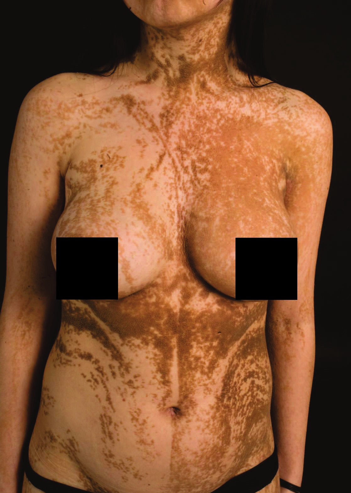 · 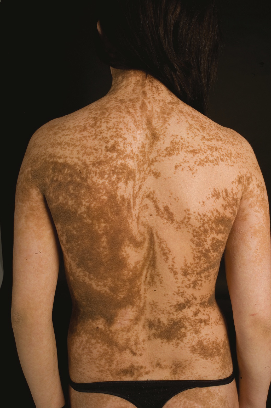 · 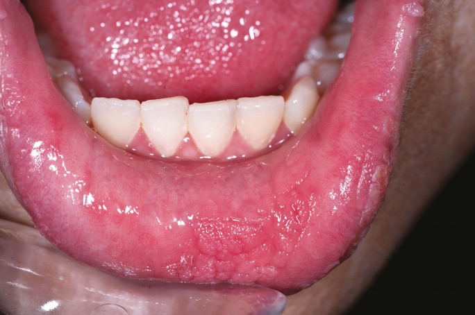 · 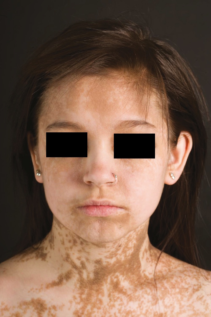 · |
|
#2
●
06-06-2025, 05:15 PM
| ||||||||
 BEHEMOTH Poster Rank:53 Blue-Eyes White Dragon Join Date: Apr 2021 Posts: 27,692
Contributions: 21
Mentioned: 322 Post(s) Quoted: 13282 Post(s)
| ||||||||
|
Re: Systemic Epidermal Nevus Due to FGFR3 Mutation
it kinda looks like a spiral on her back 
__________________ ISRAEL ✔ - PALESTINE ✗ |
|
#3
●
06-06-2025, 06:57 PM
| ||||||||
 The Devil's Left Hand Poster Rank:70 Male Join Date: May 2012 Posts: 22,189 Mentioned: 19 Post(s) Quoted: 9550 Post(s)
| ||||||||
|
Re: Systemic Epidermal Nevus Due to FGFR3 Mutation
Or like someone spilled a couple of lattes all over her. 
__________________ Why so sad? Your tears are no match for mine. |
|
#4
●
06-06-2025, 07:09 PM
| ||||||||
 My Rank: FIRST LIEUTENANT Poster Rank:244 All hail Andrés Manuel López Obrador (AMLO) Join Date: Oct 2021 Posts: 5,627
Contributions: 6
Mentioned: 186 Post(s) Quoted: 2295 Post(s)
| ||||||||
|
Re: Systemic Epidermal Nevus Due to FGFR3 Mutation
very good post, thank you
|
|
#6
●
06-07-2025, 12:51 AM
| ||||||||
 ★ Legacy Member ★ Poster Rank:118 Secret Agent Join Date: Dec 2009 Posts: 13,712 Mentioned: 6 Post(s) Quoted: 2867 Post(s)
| ||||||||
|
Re: Systemic Epidermal Nevus Due to FGFR3 Mutation
I’m sure she’s really self conscious about her condition but personally I think it’s cool looking. Something different than the norm of a single color pigment. I once saw a young woman with freckles from hell and I don’t think I’ve ever seen a more beautiful woman in my life. This is similar to me.  Everything in life is perspective. Thanks for such an informative post. |
|
#9
●
06-08-2025, 05:21 AM
| ||||||||
 ♚ Legacy Gold Member ♚ Poster Rank:267 ꓕSIꓤIꓕ∀S ϽINOᗡꓤ∀S Join Date: Feb 2010 Posts: 5,125
Contributions: 6
Mentioned: 40 Post(s) Quoted: 1425 Post(s)
| ||||||||
|
Re: Systemic Epidermal Nevus Due to FGFR3 Mutation
Clearly, God has called her to be a barista...
__________________ 200 Additional Smilies Custom DR Search Engine✦ Live life to it's fullest and leave a sexy corpse ✦ |