|
#1
●
12-21-2010, 12:37 PM
| ||||||||
| My Rank: FIRST SERGEANT Poster Rank:459 Kitty Join Date: Jul 2010 Posts: 2,369 Mentioned: 1 Post(s) Quoted: 5 Post(s)
| ||||||||
|
Lion Face Syndrome
Sorry this is long, but it is rather interesting. Leontiasis Ossea, also known as leontiasis or lion face, is a rare medical condition, characterized by an overgrowth of the facial and cranial bones. It is not a disease in itself, but a symptom of other diseases, including Paget’s disease, fibrous dysplasia, hyperparathyroidism and renal osteodystrophy, and is also seen in patients who have advanced lepromatous leprosy. A 25 year old Palestinian patient attended the author’s clinic complaining of a huge bony growth in the facial bones, orbits, nose, mandible and skull (Figs. 1, 2). At age sixteen, she underwent a minor surgery to excise left sided zygomatic bone growth through an intra-oral approach at Hadassah Hospital. She was diagnosed according to the pathology tests as having Fibrous Dysplasia and was told by the surgeons that this growth will re-occur and at age 25 further surgery may be required after cessation of the skeletal growth. Background Fibrous Dysplasia is a developmental abnormality of unknown cause in which osteoblasts fail to differentiate and mature normally. It may affect a single or many bones. Head and neck Fibrous Dysplasia is a condition in which normal medullary bone is replaced by an abnormal proliferation of fibrous tissue, resulting in asymmetrical distortion and expansion of bone; it may be confined to a single bone (monostotic fibrous dysplasia) or may involve multiple bones (polyostotic fibrous dysplasia). The monostotic form may involve any of the facial bones, but is most commonly seen in the maxilla. In the maxilla it usually presents with swelling and deformity of the cheek, but sometimes it causes nasal obstruction and/or orbital symptoms. Extensive involvement of the face is referred to as leontiasis ossea. Fibrous dysplasia is a disease of young patients. Usually no new lesions appear after the cessation of skeletal growth. The lesions become more sclerotic with time but may continue to grow slowly into adulthood. Occasionally, reactivation of the lesions occurs during pregnancy. Most patients have monostotic lesions, predominating in one of the ribs or the femur, tibia, gnathic bone, calvaria or humerus; polyostotic fibrous dysplasia more frequently involves the skull and facial bones, pelvis, spine and shoulder girdle. Radiologically, Fibrous Dysplasia appears as enlarged bone with a dense ground-glass appearance. Sometimes the lesions have a more osteolytic appearance, with regions of denser calcifications within them. Radiographs show radiolucent or sclerotic lesions, with osseous expansion almost always occurring in an outward direction. The outer table of the cranial vault is convex, and a localized zone of relative radiolucency surrounded by a sclerotic rim may be seen (doughnut lesion). Case Description Recently, the patient was seen by her surgeons at Hadassah Hospital who refused to carry out surgery due to high risk morbidity and mortality expected because the growth is so huge and highly vascular. She was indeed suffering from a rare form of Fibrous Dysplasia with slow but constant abnormal bone formation affecting and deforming all the cranial bones as well as those of the face. This condition was diagnosed by the author as Lion Face Syndrome. There is no treatment other than exposing the overgrown bone, and chipping away pieces, or excising entirely where possible. Besides enhancing the physical and psychological condition of the patient, surgical treatment was carried out by the author to prevent the senses being lost one by one and possible death finally resulting from cerebral pressure. Surgery was successfully carried out through three large surgical flaps, an apron neck incision to expose the deformed mandible and both condyles (Figs. 3, 4), a bi-coronal incision to expose the deformed frontal bone, zygoma and skull (Figs. 5-7) and an intra-oral incision to expose the deformed maxillary bone. (Figs. 8, 9) show the combined apron and bi-coronal flaps. The surgery ended up with removal of the excessive abnormal bone (Fig. 10) and re-shaping of the face and head (Figs. 11, 12). In compliance with the patient’s will not to publish her pre and post-operative personal photos, only the surgical and pre-operative and immediate post-operative radiographic photos, that are descriptive of the condition, were published. Figure 1: Preoperative lateral view Figure 2: Preoperative frontal view Figure 3: Apron neck degloving flap - Frontal view Figure 4: Apron neck degloving flap - Lateral view Figure 5: Bicoronal flap Figure 6: Bicoronal flap Figure 7: Bicoronal flap Figure 8: Apron flap & Bicoronal flap - Frontal view Figure 9: Apron flap & Bicoronal flap - Lateral view Figure 10: Excessive abnormal bone Figure 11: Immediate postoperative frontal view Figure 12: Immediate postoperative lateral view 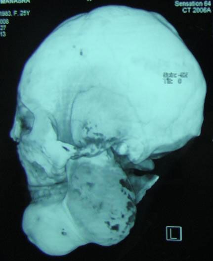 ·  · 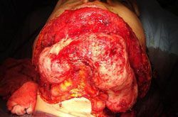 · 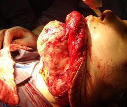 · 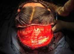 · 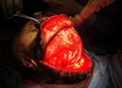 · 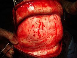 · 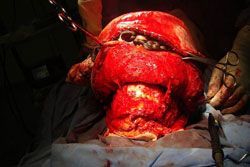 · 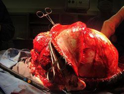 · 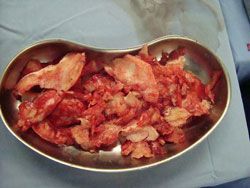 ·  ·  · |
|
#3
●
12-21-2010, 07:35 PM
| ||||||||
| ★ realist-absurdist ★ Poster Rank:138 female Join Date: Feb 2010 Posts: 11,717 Mentioned: 0 Post(s) Quoted: 16 Post(s)
| ||||||||
|
Re: Lion Face Syndrome
I agree that Cher was a good actress in that movie. I must also admit that I cried when her boy died...
|
|
#4
●
12-22-2010, 07:18 AM
| ||||||||
| So Fucking Banned Poster Rank:308 Male Join Date: Mar 2010 Posts: 4,168 Mentioned: 0 Post(s) Quoted: 0 Post(s)
| ||||||||
|
Re: Lion Face Syndrome
I am man enough to admit that I cried too. Eric Stoltz was great in the movie as his outlook throughout the whole ordeal was incredible. When she went in to wake him up and he was dead was a tough one. |
|
#6
●
12-22-2010, 02:20 PM
| ||||||||
| ★ Legacy Member ★ Poster Rank:6 What we've got here is failure to communicate. Join Date: Aug 2009 Posts: 103,591
Contributions: 3
Mentioned: 111 Post(s) Quoted: 43397 Post(s)
| ||||||||
|
Re: Lion Face Syndrome
Cher has had more plastic surgery than that freak M. Jackson, so playing the part of mum of a disfigured young one is a bit hypocritical in my opinion.
|