|
#1
●
09-30-2012, 12:24 PM
| ||||||||
| My Rank: MAJOR Poster Rank:11 Join Date: Jun 2009 Posts: 93,130
Contributions: 226
Mentioned: 91 Post(s) Quoted: 2009 Post(s)
| ||||||||
|
Post-mortem/Dissection Photos With Info
A body following skin and bone removal for transplantation purposes: 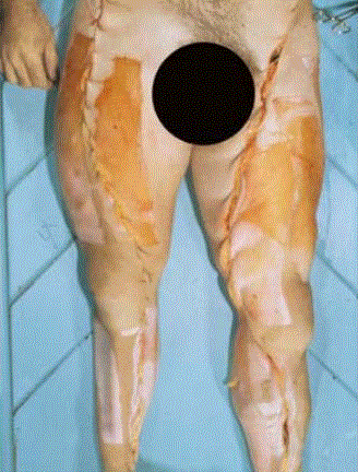 A case of meningitis, characterized by a purulent exudate (pus) covering the brain. The pus is the result of a bacterial infection: 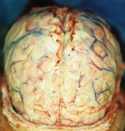 An enlarged heart from obesity or high blood pressure may be twice the size of a normal heart: 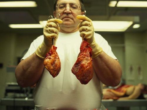 Dissection of the heart during an autopsy. The chordae tendineae, or heart strings, can be seen clearly: 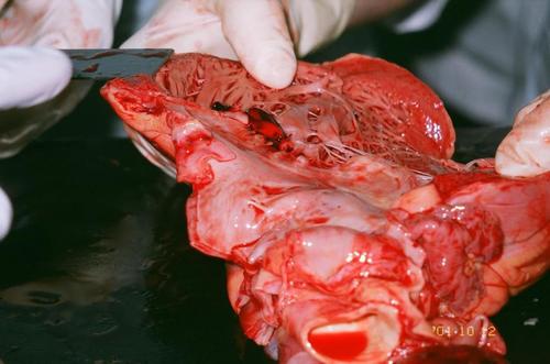 During an autopsy the brain is examined and is usually serially sectioned in the “fresh” state, although in some cases the brain may be fixed in formalin prior to sectioning, as fixationg considerably improves the ability to dissect brain tissue: 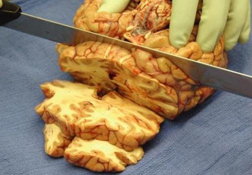 A cross section of the posterior part of the brain, the cerebellum: 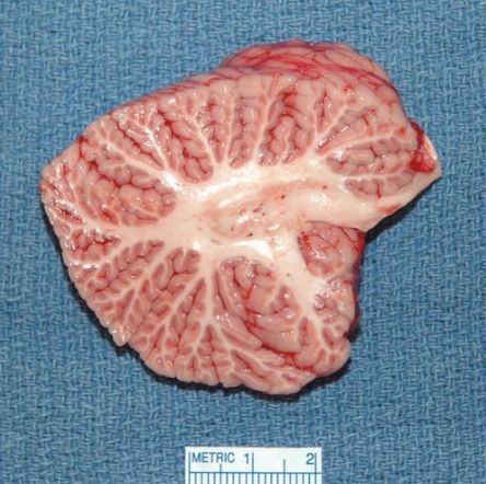 A lung showing centrilobular emphysema characteristic of smoking. Closeup of fixed, cut surface shows multiple cavities lined by heavy black carbon deposits: 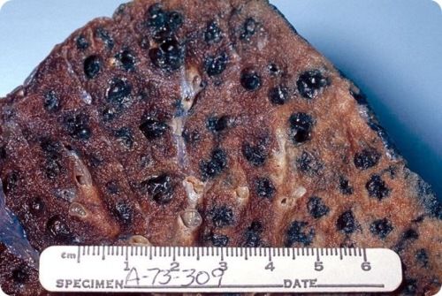 The “Y” incision is closed post autopsy, and the body is released to a funeral home. Top left is the neck, presenting a ligature mark: 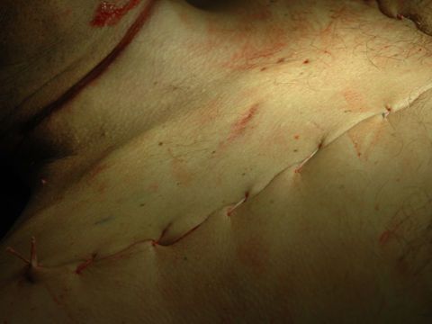 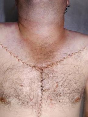 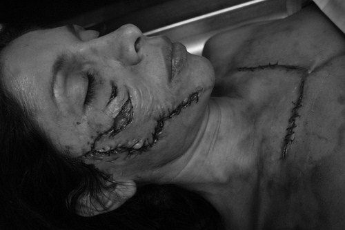 After the skullcap and dura have been removed, the brain is exposed and ready for removal from the cranial cavity: 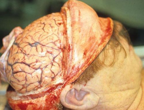 A photograph taken at autopsy following the removal of the heart, lungs, diaphragm, liver, omentum, large intestine, and a majority of the small intestines. Remaining intact are the esophagus (white arrow), the stomach, which is distended by gas, the duodenum (hidden from view), and the pancreas (the head of the pancreas is denoted by the black arrow): 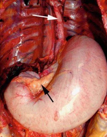 Traumatic rupture of the heart from a gunshot wound. The apex of the heart has been torn away by the bullet. The shooting was suicidal and the muzzle of the gun was held against the chest wall: 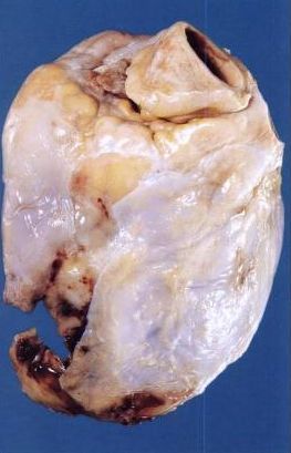 Subdural Hemorrhage - Victim thrown from a moving car:  A gunshot to the head causes a marked expansion of the brain material as the bullet travels through the brain: 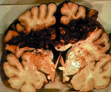 |
|
#2
●
09-30-2012, 12:44 PM
| ||||||||
| ★Wizard Of Gore★ Poster Rank:1 Join Date: Aug 2009 Posts: 126,330
Contributions: 1
Mentioned: 445 Post(s) Quoted: 30201 Post(s)
| ||||||||
|
Re: Post-mortem/Dissection Photos With Info
I would like a cafe espresso with a shot of vanilla and a purulent exudate, please. |
|
#3
●
09-30-2012, 01:49 PM
| ||||||||
| ★ Legacy Member ★ Poster Rank:2111 Male Join Date: Nov 2009 Posts: 228 Mentioned: 0 Post(s) Quoted: 16 Post(s)
| ||||||||
|
Re: Post-mortem/Dissection Photos With Info
great work Kelly I dont know how you do it
|
|
#5
●
09-30-2012, 02:24 PM
| ||||||||
| ♚ Legacy Gold Member ♚ Poster Rank:34 Female Join Date: Nov 2008 Posts: 43,403
Contributions: 204
Mentioned: 95 Post(s) Quoted: 2281 Post(s)
| ||||||||
|
Re: Post-mortem/Dissection Photos With Info
Reminds me of my gory glory years Amazing thread |
|
#6
●
09-30-2012, 02:49 PM
| ||||||||
| My Rank: LANCE CORPORAL Poster Rank:2501 Join Date: Sep 2012 Posts: 174 Mentioned: 0 Post(s) Quoted: 43 Post(s)
| ||||||||
|
Re: Post-mortem/Dissection Photos With Info
So fascinating and informative! Thank you for your excellent post!
|
|
#9
●
09-30-2012, 04:55 PM
| ||||||||
| My Rank: CORPORAL Poster Rank:1541 i am with penis Join Date: Aug 2009 Posts: 366 Mentioned: 0 Post(s) Quoted: 0 Post(s)
| ||||||||
|
Re: Post-mortem/Dissection Photos With Info
Very very cool,informative and thoughtful post, thank you !
|
|
#10
●
09-30-2012, 05:18 PM
| ||||||||
| ★The Queen★ Poster Rank:67 Female Join Date: Jun 2011 Posts: 23,498
Contributions: 120
Mentioned: 95 Post(s) Quoted: 3083 Post(s)
| ||||||||
|
Re: Post-mortem/Dissection Photos With Info
Very cool indeed! I like the presentation of the hearts. I'm glad my lungs will be pink and glistening. |