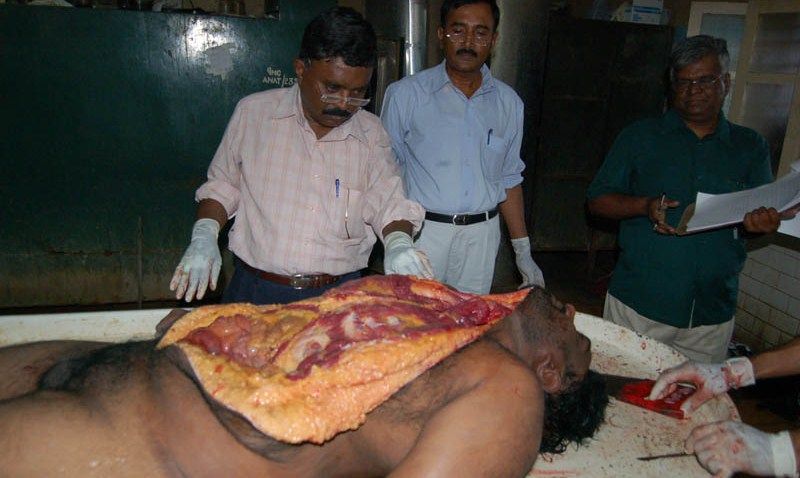
The victim was a middle aged man, apparently beaten to death.
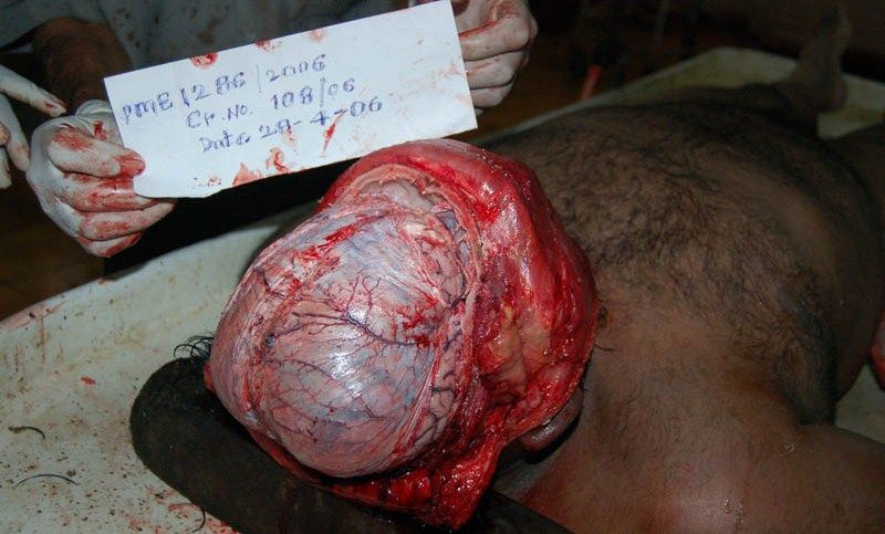
1-In this first image we can see that they have already taken off the top of his skull revealing the brain and brain sac.
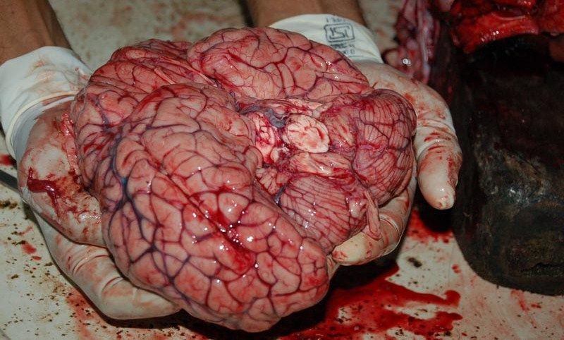
2-Here we see the brain freed from the brain pan.
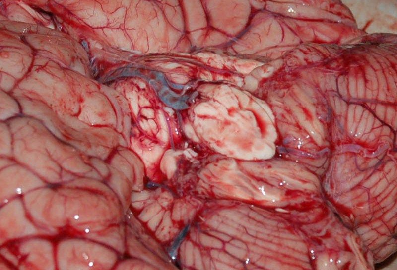
3-In this close up of the previous picture we can clearly see the Medulla Oblongata which is the lower portion of the brain stem and is continuous with the spinal cord. Prior to brain removal it is normal procedure for the pathologist to take a sample of cerebrospinal fluid Using a wide bore pipette passed gently over the front of the Medulla Oblongata prior to it being severed to remove the brain.
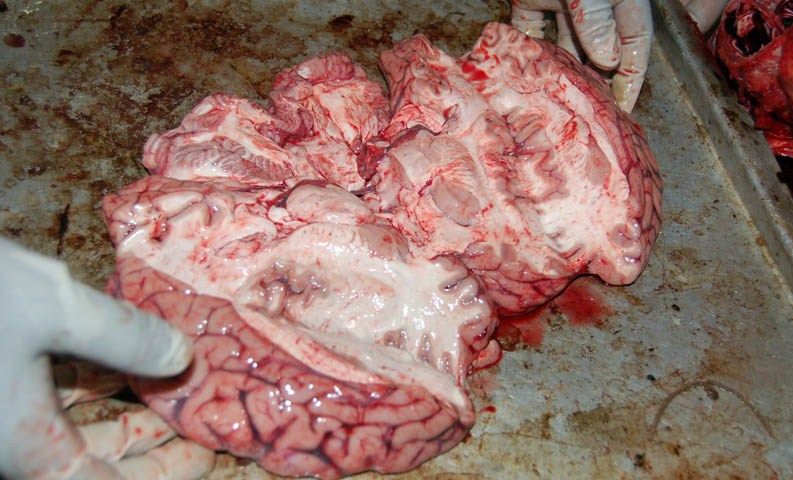
4-The pathologist would, after a visual examination of the brain exterior, dissect the brain to check for damage to the interior. It is here where haemorrhages become readilly apparent.
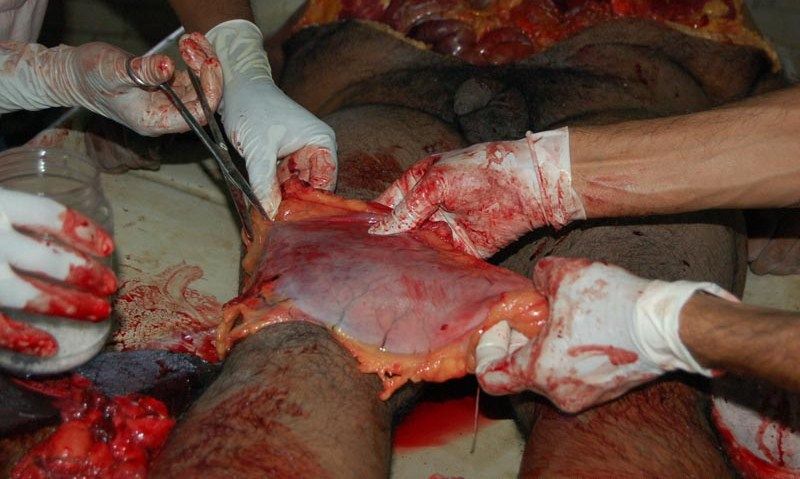
5-Now the pathologist begins on the dissection of the torso. The incisions made often follow a set method.The usual incision starts behind the ears then runs along the neck to the clavicle (Also known as your collar bone). A traverse cut across the manubrium sterni (Breastbone) joins the lateral neck incisions. This is followed by a straight midline thoraco-abdominal cut which runs down the chest and stomach. The neck flap is lifted towards the head and the chest-stomach flaps are directed laterally. In this case it would appear the pathologist has simply cut straight from the throat down to the stomach
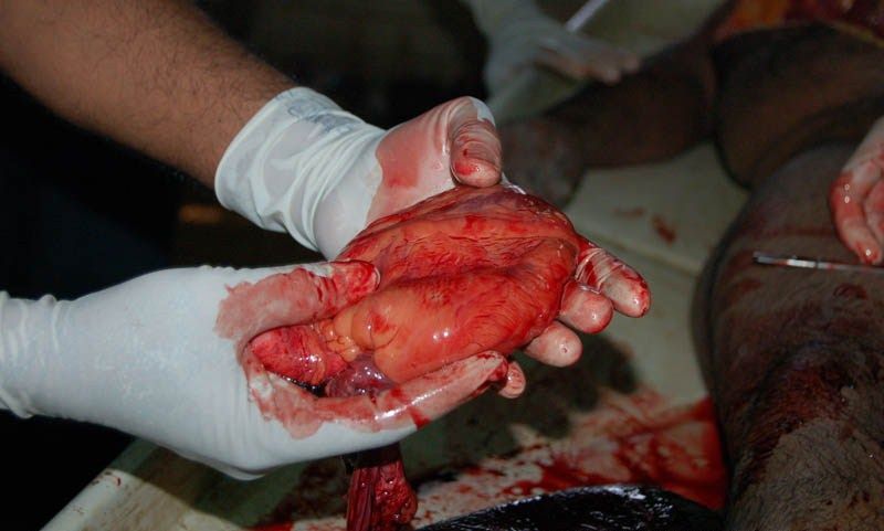
6-Here we see the heart taken from the victims chest. As with the brain a careful examination of the organs exterior is carried out.
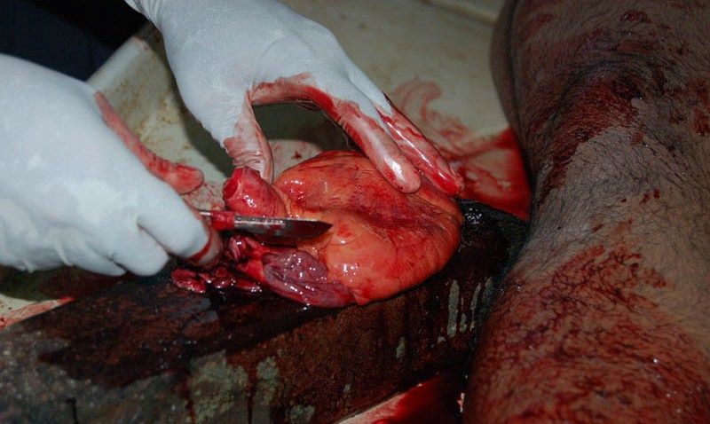
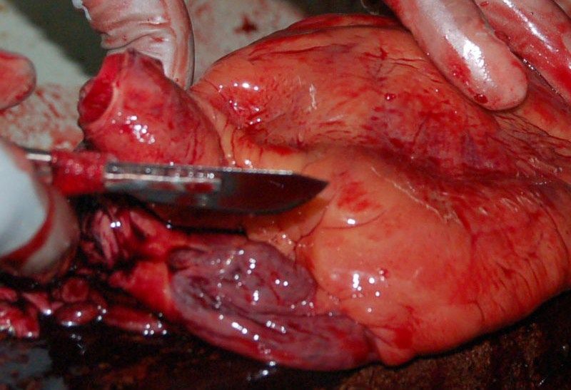
7 and 8-Prior to dissection.
9-All organs are removed from the body, examined, and often weighed.
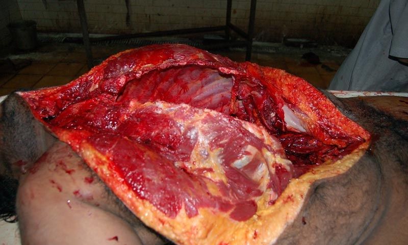
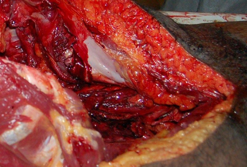
10-Here we can see the, now empty, chest and stomach cavities.
11-And finally a close up detailing the emptied stomach cavity clearly showing the fatty layer beneath the skin of the victims stomach.


