|
#1
●
12-07-2008, 03:31 PM
| ||||||||
| My Rank: FIRST SERGEANT Poster Rank:445 DR's Poptart Join Date: Nov 2008 Posts: 2,517 Mentioned: 1 Post(s) Quoted: 26 Post(s)
| ||||||||
|
Pictures of a 50-year-old Woman With Mammary Sarcomas
Whatever it is, it's freakin' gross! 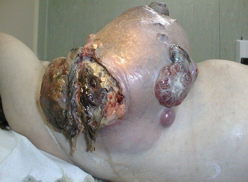 ·  ·  ·  · 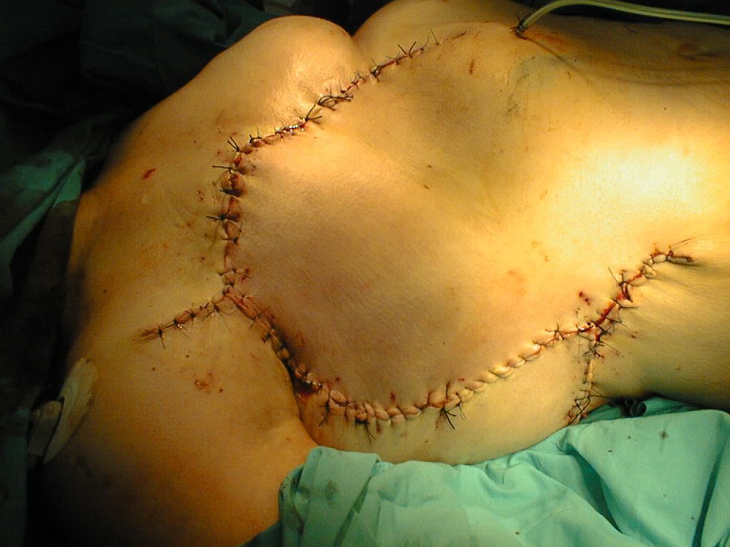 · 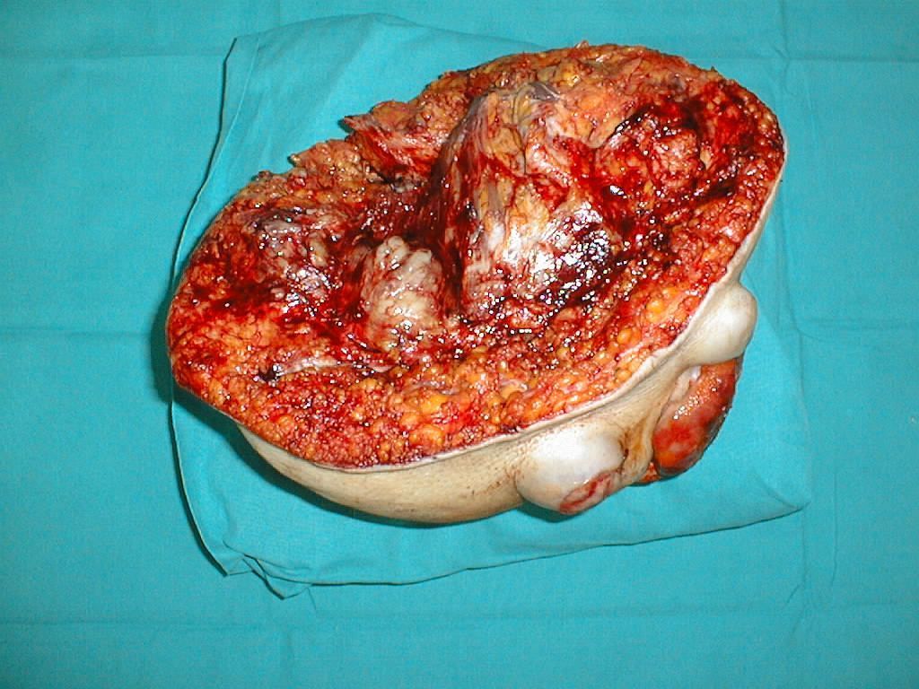 · 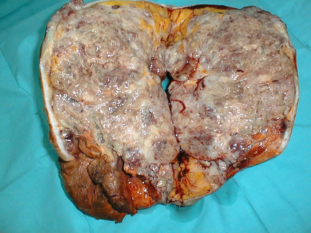 ·  · |
|
#2
●
12-07-2008, 04:02 PM
| ||||||||
| ★ ★ GENERAL ★ ★ Poster Rank:42 male Join Date: Oct 2006 Posts: 34,450
Contributions: 35
Mentioned: 2166 Post(s) Quoted: 20744 Post(s)
| ||||||||
|
Re: Pictures of a 50-year-old Woman With Mammary Sarcomas
wait a second, .... That's some woman's tit!
__________________ Support the site and our continued existance by upgrading to a Green Name |
|
#4
●
12-07-2008, 05:36 PM
| ||||||||
| ★ ★ GENERAL ★ ★ Poster Rank:42 male Join Date: Oct 2006 Posts: 34,450
Contributions: 35
Mentioned: 2166 Post(s) Quoted: 20744 Post(s)
| ||||||||
|
Re: Pictures of a 50-year-old Woman With Mammary Sarcomas
I don't know, kinda looks like it
__________________ Support the site and our continued existance by upgrading to a Green Name |
|
#5
●
12-07-2008, 05:40 PM
| ||||||||
| My Rank: FIRST SERGEANT Poster Rank:445 DR's Poptart Join Date: Nov 2008 Posts: 2,517 Mentioned: 1 Post(s) Quoted: 26 Post(s)
| ||||||||
|
Re: Pictures of a 50-year-old Woman With Mammary Sarcomas
I think your right! It's totally a tit! What the hell is wrong with it though?? |
|
#10
●
12-08-2008, 10:40 PM
| ||||||||
| Bananananananana Poster Rank:554 Lass Join Date: Dec 2008 Posts: 1,770 Mentioned: 0 Post(s) Quoted: 8 Post(s)
| ||||||||
|
Re: Pictures of a 50-year-old Woman With Mammary Sarcomas
Really? I usually just cuss at them for being in the way |